
|
Fig. 5
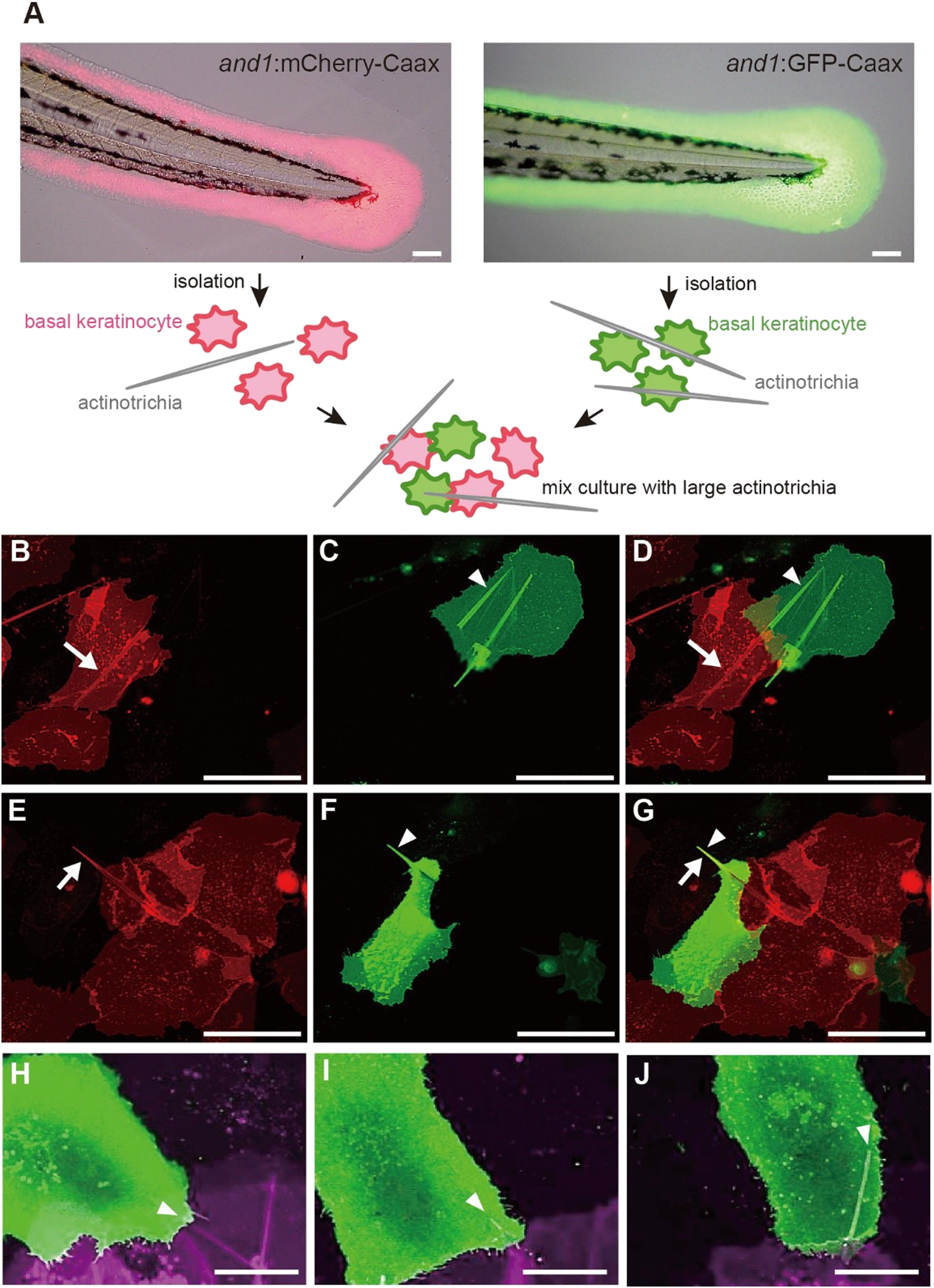
In vitro holding of the long mature actinotrichia by the basal keratinocytes from two reporter lines. (A) Schematic diagram of the mix-culture experiment using two types of reporter zebrafish. Scale bar is 50 μm. (B–J) The cultured basal keratinocytes in vitro from the larvae of and1:mCherry-CaaX and and1:GFP-CaaX. (B) White arrow indicates the actinotrichium in the basal keratinocyte expressing mCherry-Caax. (C) White arrowhead indicates the actinotrichium in the basal keratinocyte expressing GFP-CaaX. (E) White arrow indicates the actinotrichium protruded from a basal keratinocyte were enveloped by a mCherry-CaaX-expressing basal keratinocyte. (F) White arrowhead indicates GFP-CaaX-expressing basal keratinocyte. (D and G) Merged image of (B and C) and (E and F), respectively. Two adjacent basal keratinocytes share the same actinotrichia. Scale bar in (B − G) is 50 μm. (H–J) Time-lapse images of the plasma membrane enveloping the actinotrichium. White arrowheads indicate the actinotrichium enveloped by two adjacent basal keratinocytes. Scale bar in (H–J) is 20 μm.
Reprinted from Mechanisms of Development, 153, Kuroda, J., Iwane, A.H., Kondo, S., Roles of basal keratinocytes in actinotrichia formation, 54-63, Copyright (2018) with permission from Elsevier. Full text @ Mech. Dev.