
Fig. S3
- ID
- ZDB-IMAGE-190814-5
- Publication
- Lin et al., 2019 - An Ectoderm-Derived Myeloid-like Cell Population Functions as Antigen Transporters for Langerhans Cells in Zebrafish Epidermis
- All Figures
- Figures for Lin et al., 2019
|
Fig. S3
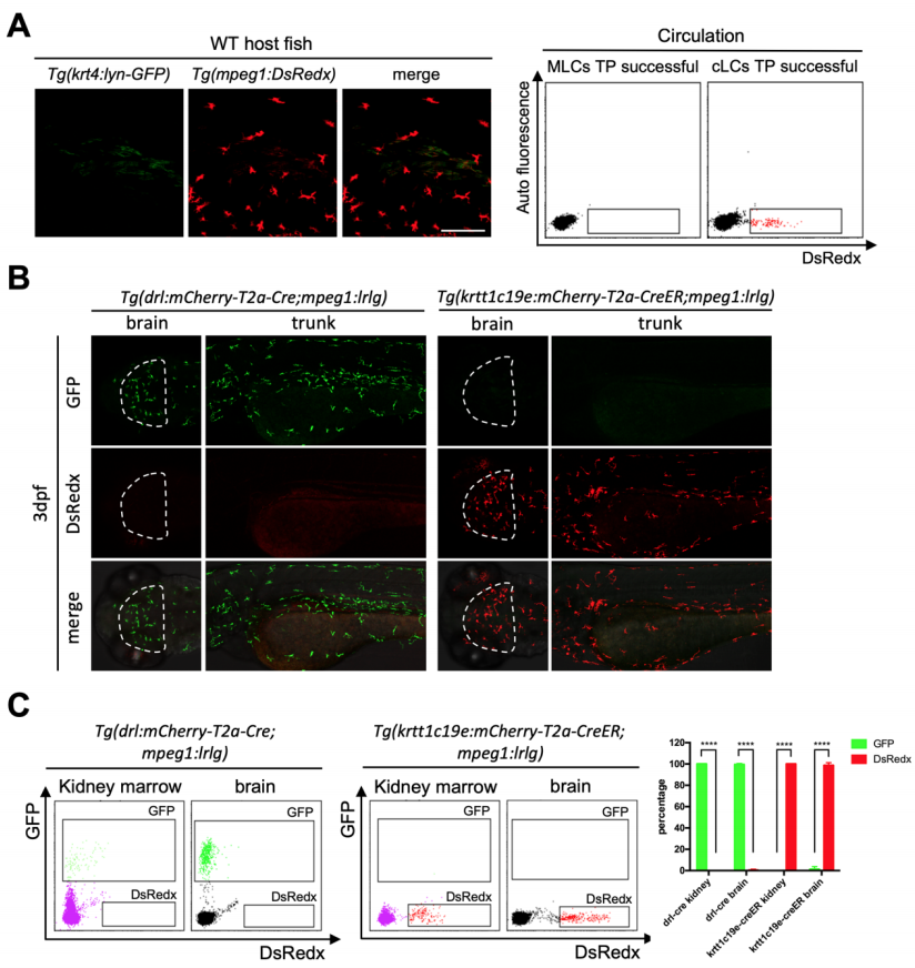
Tg(drl:mCherry-T2a-Cre) and Tg(krtt1c19e:mCherry-T2aCreERT2) fish specifically and efficiently label mesoderm derived blood cells and ectoderm derived cells respectively. Related to Figure 3 (A) Successful transplantation of cLCs in 2 months old recipients (n=5). Donor mpeg1+ cells (red) (representing cLCs) widely distribute throughout whole fish epidermis while no krt4+ cells (green) exist. Scale bar, 100μm. FACS data show that only cLCs successful transplanted fish contain DsRedx+ cells in circulation. (B) Confocal images of brain (white dash lines) and trunk region of Tg(drl:mCherry-T2a-Cre;mpeg1:loxP-DsRedx-loxP-GFP) and Tg(krtt1c19e:mCherry-T2a-CreERT2;mpeg1:loxP-DsRedx-loxP-GFP) (add 4- OHT 1-5dpf) fish at 3dpf (each group, n=8 fish). All microglia and trunk macrophage are labeled by mesodermal line while no cells could be labeled by ectodermal line. Scale bar, 200μm. (C) FACS analysis of kidney marrow and brain mpeg1+ cells in adult fish. Nearly 100% mpeg1+ cells in Tg(drl:mCherry-T2a-Cre;mpeg1:loxP-DsRedx-loxP-GFP) fish are GFP+, while no cells in Tg(krtt1c19e:mCherry-T2aCreERT2;mpeg1:loxP-DsRedx-loxP-GFP) (add 4-OHT 1-5dpf) fish are GFP+. Quantification of GFP+ and DsRedx+ cells percentages to total mpeg1+ population in kidney marrow and brain (each group, n=4). Data are represented as mean ± SD, ****P<0.0001.
Reprinted from Developmental Cell, 49(4), Lin, X., Zhou, Q., Zhao, C., Lin, G., Xu, J., Wen, Z., An Ectoderm-Derived Myeloid-like Cell Population Functions as Antigen Transporters for Langerhans Cells in Zebrafish Epidermis, 605-617.e5, Copyright (2019) with permission from Elsevier. Full text @ Dev. Cell