
Fig. 3
- ID
- ZDB-IMAGE-190813-10
- Publication
- Lin et al., 2019 - An Ectoderm-Derived Myeloid-like Cell Population Functions as Antigen Transporters for Langerhans Cells in Zebrafish Epidermis
- All Figures
- Figures for Lin et al., 2019
|
Fig. 3
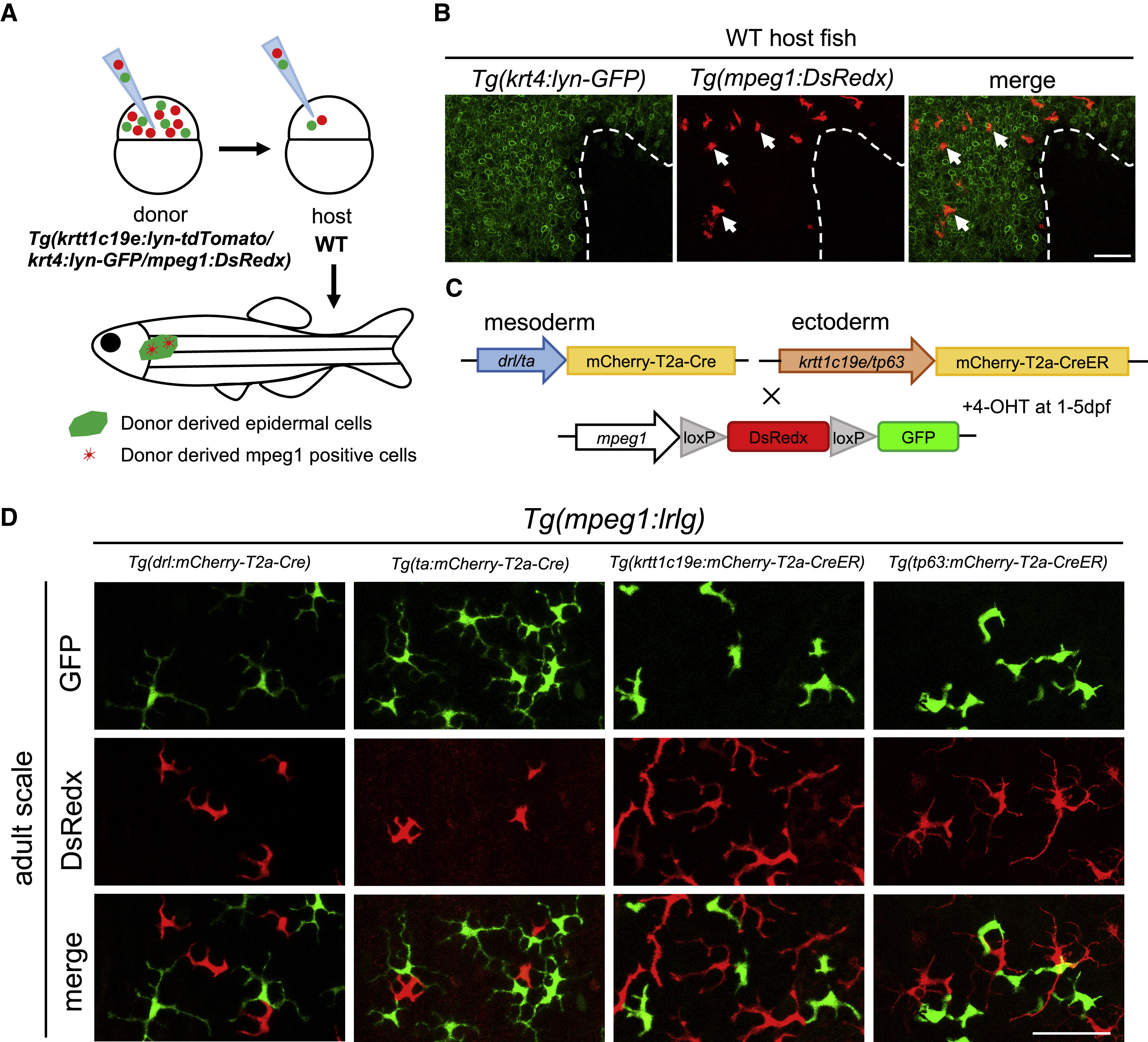
MLCs Are Derived from Ectodermal Origin
(A) A schematic draw of cell transplantation assay. About 30 cells from Tg(krtt1c19e:lyn-tdTomato;krt4:lyn-GFP;mpeg1:DsRedx) donor embryos are collected at 3.5 hpf and transplanted into WT embryos at the same stage. Chimeric embryos with successful contribution of donor cells to ectodermal cells (tdTomato+) are grown to 2 months old and subjected to correlation analysis for donor-derived mpeg1+ cells (DsRedx+) and keratinocytes in the epidermis (lyn-GFP+).
(B) Donor mpeg1+ (red) (representing MLCs) and krt4+ cells (green) co-exist in the epidermis of 2-month-old recipients (n = 11). White dash line indicates the mosaic boundary. Scale bar, 50 μm.
(C) A schematic diagram shows the strategy to track mesoderm- and ectoderm-derived mpeg1+ cells in the epidermis of adult fish. Mesoderm-specific Cre lines Tg(drl/ta:mCherry-T2a-Cre) and ectoderm-specific CreER lines Tg(krtt1c19e/tp63:mCherry-T2a-CreERT2) are outcrossed with Tg(mpeg1:loxP-DsRedx-loxP-GFP) reporter line. The resulting ectoderm-specific CreER-reporter double transgenic lines are then treated with 4-OHT at 1 dpf for 4 days.
(D) Confocal images of adult scale epidermis from four groups of fate-mapping double transgenic fish. Both mesoderm-specific (representing cLCs) and ectoderm-specific (representing MLCs) double transgenic fish contain GFP+ cells (each group, n = 10 fish). Scale bar, 50 μm.
Reprinted from Developmental Cell, 49(4), Lin, X., Zhou, Q., Zhao, C., Lin, G., Xu, J., Wen, Z., An Ectoderm-Derived Myeloid-like Cell Population Functions as Antigen Transporters for Langerhans Cells in Zebrafish Epidermis, 605-617.e5, Copyright (2019) with permission from Elsevier. Full text @ Dev. Cell