
IMAGE
Figure S1
- ID
- ZDB-IMAGE-190812-6
- Publication
- Ouyang et al., 2019 - CPSF1 mutations are associated with early-onset high myopia and involved in retinal ganglion cell axon projection
- All Figures
- Figures for Ouyang et al., 2019
Image
|
Figure Caption
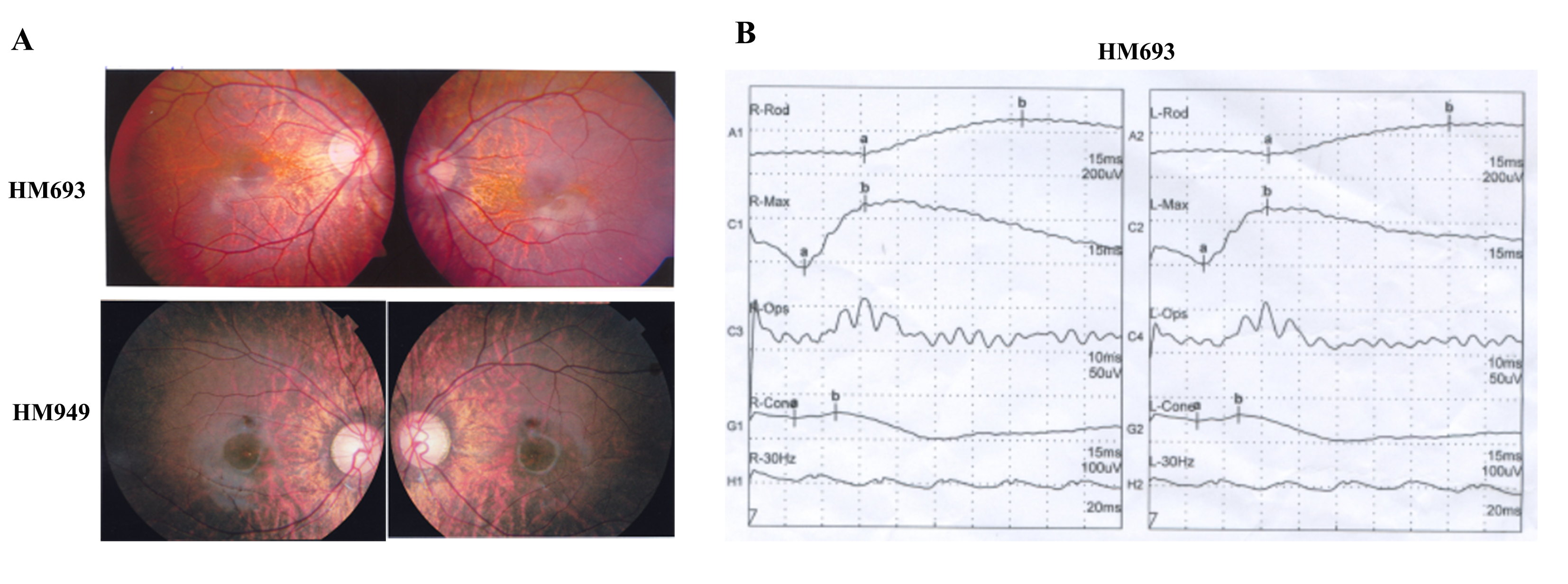
Figure S1
. Available fundus changes of the probands HM693 and HM949 and ERG recording of the proband HM693. (A) Typical fundus changes of high myopia, including an optic nerve head crescent and a “tigroid” appearance of the posterior retina. (B) ERG recording of the proband HM693 showed severely reduced cone responses and mildly reduced rod responses
Acknowledgments
This image is the copyrighted work of the attributed author or publisher, and
ZFIN has permission only to display this image to its users.
Additional permissions should be obtained from the applicable author or publisher of the image.
Full text @ Hum. Mol. Genet.