
Fig. 4
- ID
- ZDB-IMAGE-190812-23
- Genes
- Publication
- Elworthy et al., 2019 - The role of endothelial cilia in post-embryonic vascular development
- All Figures
- Figures for Elworthy et al., 2019
|
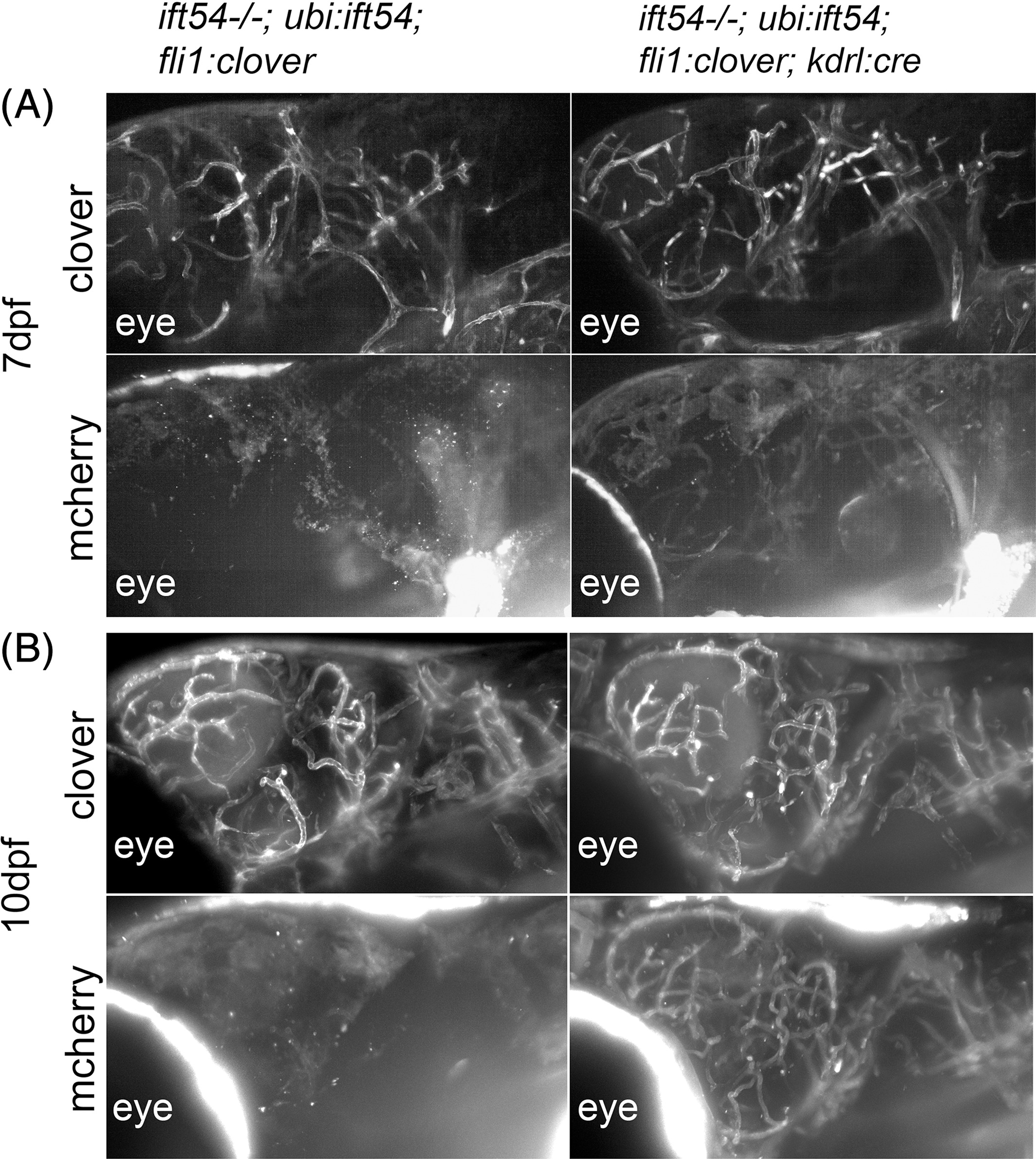
Fig. 4
Endothelial‐specific excision of the Tg(ubi:loxP‐ift54‐loxP‐myr‐mcherry,myl7:EGFP)sh488 rescue transgene. A: SPIM stack projections of lateral views of the head of 7‐dfp ift54 tp49; Tg(ubi:loxP‐ift54‐loxP‐myr‐mcherry,myl7:EGFP)sh488 /+;Tg(fli1a:LIFEACT‐clover)sh467 /+ (ift54−/−; ubi:ift54; fli1:clover) and ift54 tp49; Tg(ubi:loxP‐ift54‐loxP‐myr‐mcherry,myl7:EGFP)sh488 /+;Tg(kdrl:cre)s898/+;Tg(fli1a:LIFEACT‐clover)sh467 /+ (itf54−/−; ubi:ift54; fli1:clover; kdrl:cre) fish showing endothelial Clover and faint mcherry expression. The position of the eye is labeled for orientation. Endothelial‐specific Cre‐mediated recombination of the Tg(ubi:loxP‐ift54‐loxP‐myr‐mcherry,myl7:EGFP)sh488 transgene is marked by mcherry expression. B: At 10 dpf, mcherry expression is more pronounced than at 7 dpf