
|
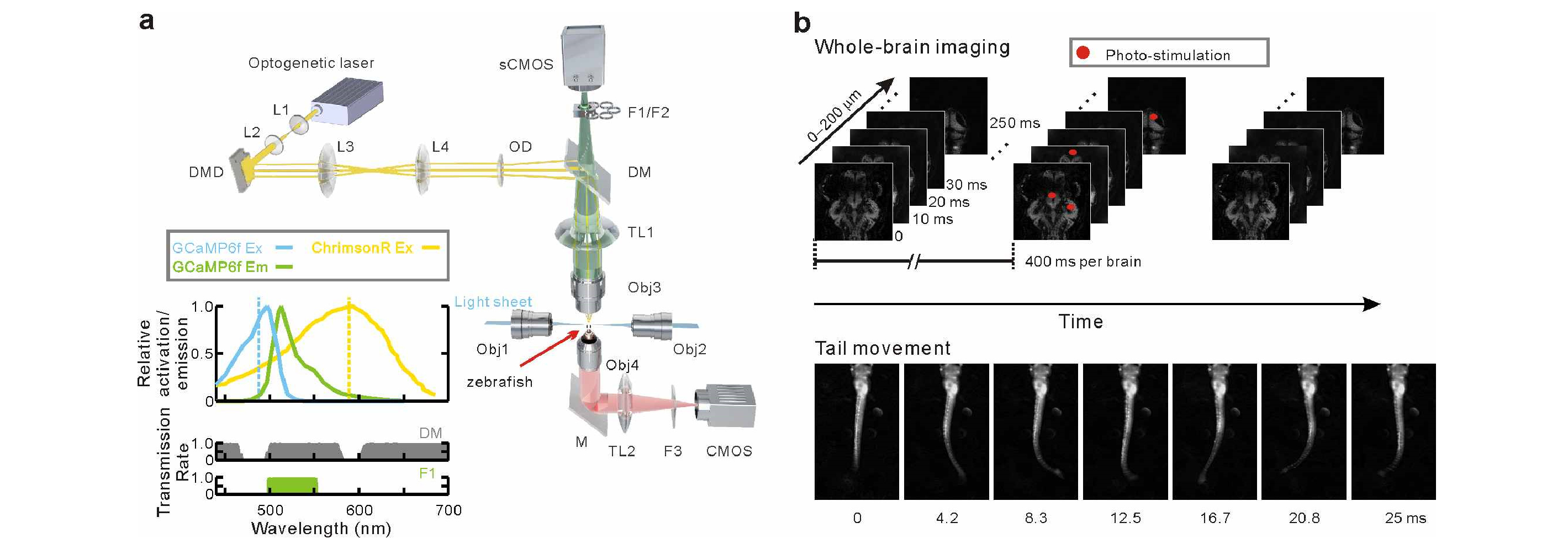
Fig. 1
Simultaneous whole-brain imaging and targeted manipulation of neuronal activity of behaving larval zebrafish. (a) Schematic diagram of setup. Patterned photo-stimulation was incorporated into a light-sheet microscope through the imaging light path, and a near-infrared light path was used for behavior monitoring. (bottom left) GCaMP6f, ChrimsonR, and filter combination spectra. The separated GCaMP6f and ChrimsonR spectra allowed simultaneous light-sheet imaging and photo-stimulation without spectral bleeding. ChrimsonR Ex: ChrimsonR excitation spectrum; GCaMP6f Ex and Em: GCaMP6f excitation and fluorescence emission spectra, respectively. Blue and yellow dashed lines: GCaMP6f (488 nm) and ChrimsonR (589 nm) excitation wavelengths, respectively. (b) Simultaneous targeted manipulation and whole-brain imaging of neuronal activity (top) and behavior monitoring (bottom). Distributed neurons were targeted with photo-stimulation (red dots) during whole-brain neuronal activity imaging and fish behavior monitoring. (DMD: digital micromirror device; L1−L4: lenses 1−4; OD: optical diffuser; (s)CMOS: (scientific) complementary metal-oxide semiconductor; F1, F2: green and red band-pass filters, respectively; F3: infrared long-pass filter; DM: multi-band dichroic mirror; TL1, TL2: tube lenses 1 and 2; Obj3, Obj4: fluorescence detection and behavior monitoring objectives, respectively; M, reflective mirror.)