
|
Fig. S1
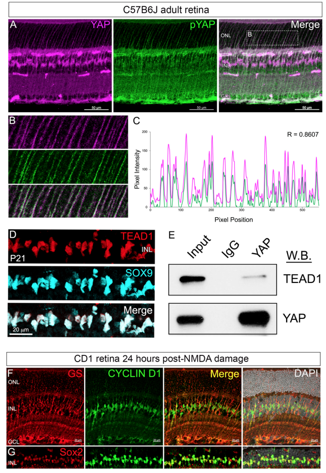
The YAP antibody labels both the MG nuclei in the INL and the cytoplasmic processes spanning the adult retina, but the pYAP antibody does not label MG nuclei (A) . In the MG cytoplasmic processes, YAP and pYAP signals overlap (B-C) . TEAD1 immunofluorescence on adult retinal cryosections showed clear localization to SOX9+ MG nuclei (D) . Whole adult retinal lysates wereimmunoprecipitated with antibodies against YAP or an IgG negative control. Western blot with anti-TEAD1 and YAP antibodies showed that YAP and TEAD1 co-immunoprecipitate (E) . By 24 hours post NMDA damage, wild type retinae exhibited a significant increase in Cyclin D1 mRNA and protein expression (see Figure 2A, 2D-F). To verify that the increase in CYCLIN D1 occurs in MGs, we performed co-immunofluorescence with antibodies against MG markers GS and SOX2 and showed that the CYCLIN D1 immunofluorescent signal is within MGs (FG) . N = 3 per sample. ONL = outer nuclear layer, INL = inner nuclear layer, GCL = ganglion cell layer.