
Fig. S1
- ID
- ZDB-IMAGE-190731-23
- Publication
- Nichols et al., 2019 - Pioneer axons employ Cajal's battering ram to enter the spinal cord
- All Figures
- Figures for Nichols et al., 2019
|
Fig. S1
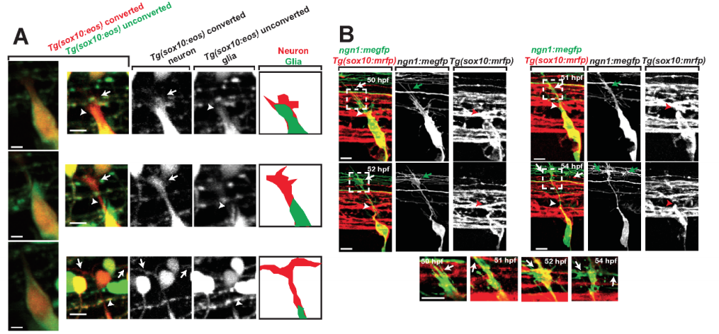
Supplementary Figure 1. Glial projections do not extend with the pioneer axon to the DREZ, related to Figure 1. (A). Confocal z-projection frames from a 24-hr timelapse starting at 48 hpf of a Tg(sox10:eos) zebrafish with a photoconverted neuron. White arrow denotes pioneer axon growth cone. White arrowhead denotes trail-ing glial process. Images on the right show the growth cone contacting the DREZ with traced representations of the neuron (red) and glia (green) (B). Confocal z-projection frames of a 24-hr time-lapse starting at 48 hpf of a Tg(sox10:mrfp) with mosaic expression of ngn1:megfp. The pioneer axon leads the associated glia throughout its navigation before, during, and after DREZ entry. White and green arrows denotes growth cone, and white and red arrowheads denotes the trailing glia. Insets shown below represent the pioneer growth cone in single-planes. Scale bars denote 10 µm.