
|
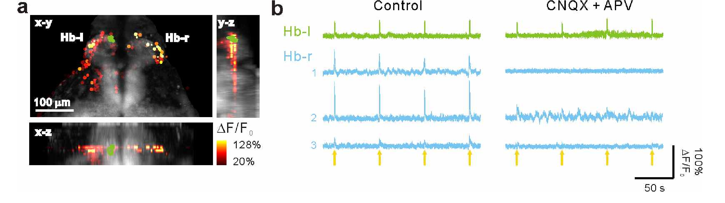
Fig. 8
Targeted photo-stimulation of habenulae evokes synaptic transmission. (a) Postsynaptic responses evoked by targeted photo-stimulation. Nine neurons (green) in the left dorsal habenula (Hb-l) and activated neurons (color dots) in bilateral Hb (55 Hb-l and 11 Hb-r (the right dorsal habenula)) in a 6-dpf Tg(elavl3:H2B-GCaMP6f;elavl3:ChrimsonR-tdTomato) larva were targeted for photo-stimulation. The neuronal activities were measured based on the amplitude of the GCaMP6f fluorescence change (ΔF/F0) and superimposed on reference anatomy (grey) in dorsal (x-y), sagittal (y-z) and coronal (x-z) projections. (b) Representative neuronal responses evoked by targeted photo-stimulation before (left) and after (right) blockade of glutamatergic transmission through bath application of CNQX (50 μM) and APV (50 μM). The green traces indicate the response of a targeted neuron in Hb-l, and the blue traces display the postsynaptic responses of three activated neurons in Hb-r. The yellow arrows indicate the targeted photo-stimulation events.