
|
Fig. 3
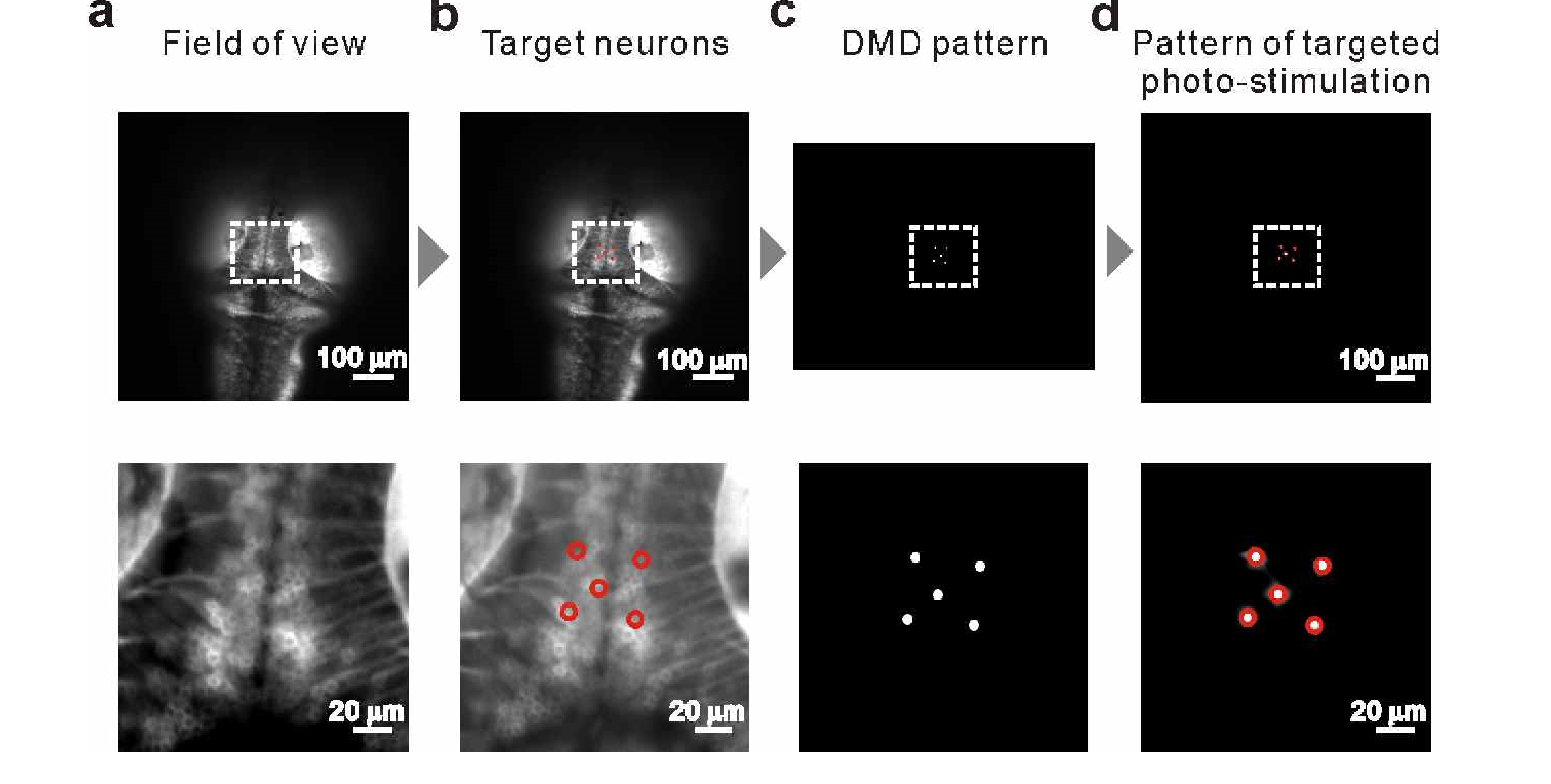
Pipeline for photo-stimulation of targeted neurons. From the captured fluorescence image of the brain of a 5-dpf zebrafish larva pan-neuronally expressing ChrimsonR-tdTomato (a), we arbitrarily selected single neurons (red circles in (b)) as photo-stimulation targets. Based on these neurons' positions and soma sizes, we calculated the photo-stimulation pattern and loaded it onto the DMD (c) to activate the target-corresponding micromirrors. Upon this targeted photo-stimulation, tdTomato fluorescence was imaged from the same field of view as for (a), and fluorescence was observed on the target neurons. (a and d, bottom) Enlarged view of region of interest (white dashed square) in each panel.