
Fig. S1
- ID
- ZDB-IMAGE-190730-8
- Publication
- Phan et al., 2019 - Differential actinodin1 regulation in embryonic development and adult fin regeneration in Danio rerio
- All Figures
- Figures for Phan et al., 2019
|
Fig. S1
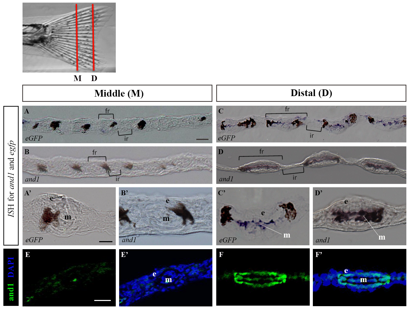
Tg(2P-EIand1:eGFP) recapitulates endogenous and1 expression in developing caudal fin at juvenile stage.
All experiments were performed on transverse cryosections of middle region (A-A’, B-B’, E, G) and distal region (C, D, F, H) of developing caudal fin of 40dpf juvenile fish. In situ hybridization (n = 8) for eGFP of Tg(2P-EIand1:eGFP) (A-A’, C, C’) and endogenous and1 expression (B-B’, D, D’). The fin rays and the interrays are indicated by brackets. (A’-D’) Higher magnification of a single fin ray from panels A-D, respectively. (E, F) Immunohistochemistry (n = 9) for And1/2. (E’, F’) merge with DAPI staining, which stains cell nuclei. n = # of fish of which fins were sectioned, and on which given experiment was performed. e: epithelium; fr: fin ray; ir: interray; m: mesenchyme. Melanocytes are the dark spots indicated by an asterisk. Scale bars: Panel A, B, C, D = 20μm, A’, B’, C’, D’ = 10μm, E-E’, F-F’ = 20μm.