
Image
|
Figure Caption
Fig. S1
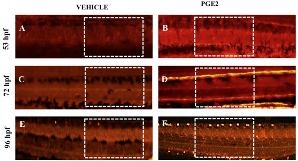
We captured vascular development of hatched zebrafish embryos at different time points 53, 72 and 96hpf with a fluorescent microscope. A region of interest (ROI) for vascular maturation was shown with white dotted rectangle boxes. Vascular development for the vehicle at three-time points are presented in A, C, E and for PGE2 presented in B, D, F.
Acknowledgments
This image is the copyrighted work of the attributed author or publisher, and
ZFIN has permission only to display this image to its users.
Additional permissions should be obtained from the applicable author or publisher of the image.
Full text @ Biol. Open