IMAGE

Figure 1 - figure supplement 2

ID
ZDB-IMAGE-190723-804
Source
Figures for Hardy et al., 2019
Image
Figure Caption

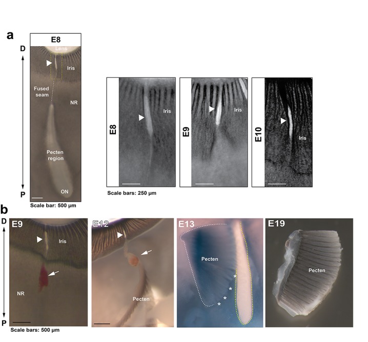
Figure 1 - figure supplement 2 Anatomical features of iris and pecten in relation to OFC in the chick eye.

(a) Iris development in flat mounted OFMs from E8-E10. Note the OFM at the iris remains open and is non-pigmented throughout all stages analysed (arrowheads). (b) Location and orientation of the developing pecten oculi and associated blood vessel (arrows) entering at the open iris region (arrowheads) shown in eyes taken from embryos at E9-19. The pecten was partially dissected from the underlying tissue (asterisks) to indicate its location relevant to the proximal optic fissure (yellow hatching). The P-D axes are shown.

Acknowledgments
This image is the copyrighted work of the attributed author or publisher, and ZFIN has permission only to display this image to its users. Additional permissions should be obtained from the applicable author or publisher of the image. Full text @ Elife