IMAGE

Fig 3

ID
ZDB-IMAGE-190723-563
Source
Figures for Kny et al., 2019
Image
Figure Caption

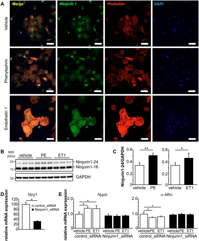
Fig 3 Knockdown of Ninjurin1 by siRNA impairs cardiomyocyte hypertrophy <italic>in vitro</italic>.

(A) Immunofluorescent staining of NRVM treated with vehicle, phenylephrine (100 μM) and endothelin 1 (50 nM) with anti-Ninjurin1 as primary antibody and Alexa Fluor 488 conjugated secondary antibody (green). Nuclei were stained with DAPI (blue). Scale bar, 75 μm. (B) Immunoblots (left) and densitometric analysis (right) of proteins isolated from vehicle, phenylephrine (100 μM) and endothelin 1 (50 nM) treated NRVM using anti-Ninjurin1 antibody. GAPDH was used as loading control. The 16kDa (Ninjurin1-16) and 24kDa (Ninjurin1-24) Ninjurin1 isoforms are indicated. (C) Densitometric analysis of the Ninjurin1-24 signal from (B). (D, E) NRVM were transfected with control siRNA (control_siRNA) (n = 3) or siRNA targeting Ninjurin1 (Ninjurin1_siRNA) (n = 3). (D) Quantitative RT-PCR analysis of Ninjurin1 (Ninj1). GAPDH expression was used as reference. Data are presented as mean ± SD. * P < 0.05. (E) Quantitative RT-PCR analysis of Nppb and alpha-myosin heavy chain 1 (α-Mhc). GAPDH expression was used as reference. Data are presented as mean ± SD. * P < 0.05, ** P < 0.01, *** P < 0.001.

Acknowledgments
This image is the copyrighted work of the attributed author or publisher, and ZFIN has permission only to display this image to its users. Additional permissions should be obtained from the applicable author or publisher of the image. Full text @ PLoS One