IMAGE

Figure 3

ID
ZDB-IMAGE-190723-452
Source
Figures for Carnovali et al., 2019
Image
Figure Caption

Figure 3

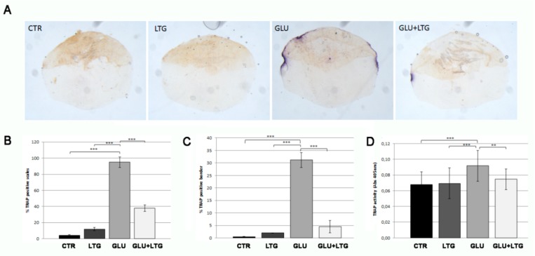
(A) Histological TRAP activity assay. Enzymatic activity along scale border of CTR scales or treated with LTG, GLU, and GLU + LTG can be visualized by violet staining. (B) Percentage of TRAP-positive scales (CTR vs. GLU, p < 0.001) (GLU + LTG vs. GLU, p < 0.001). (C) TRAP-activity extension calculation among TRAP-positive scales, (CTR vs. GLU, p < 0.001) (GLU + LTG vs. GLU, p < 0.001). (D) Biochemical TRAP assay. Quantification of TRAP activity was performed on same samples with a biochemical assay (CTR vs. GLU, p < 0.001; GLU + LTG vs. GLU, p < 0.001). p < 0.01 (**) and p < 0.001 (***).

Acknowledgments
This image is the copyrighted work of the attributed author or publisher, and ZFIN has permission only to display this image to its users. Additional permissions should be obtained from the applicable author or publisher of the image. Full text @ Nutrients