IMAGE

Fig. 1.

ID
ZDB-IMAGE-190723-2727
Source
Figures for Turcotte et al., 2019
Image
Figure Caption

Fig. 1.

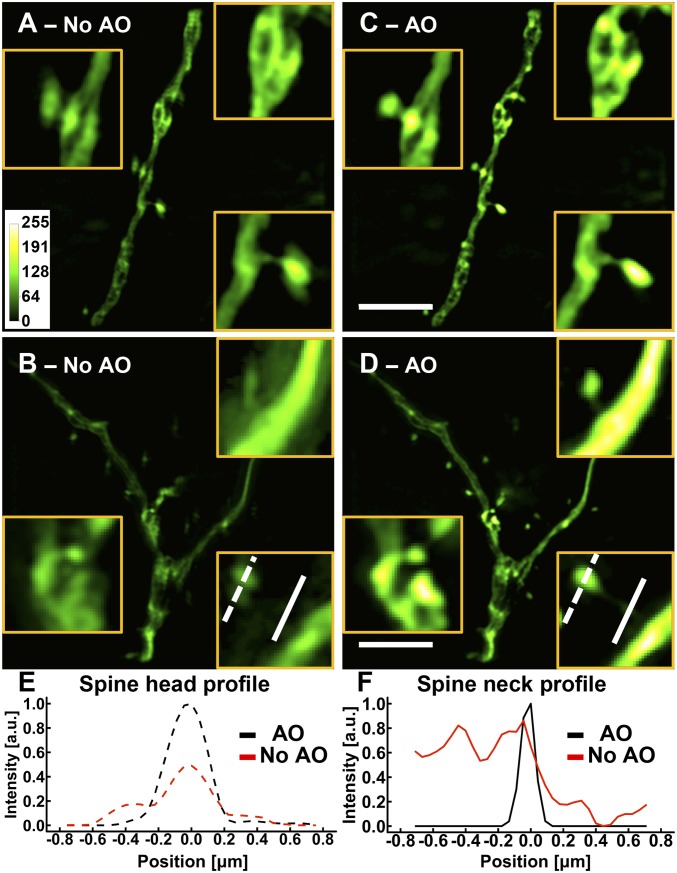
AO is essential for SIM imaging in brain tissue. (AD) Images of dendrites at a depth of 25 μm in a cortical slice of a Thy1-GFP line M mouse (A and B) without and (C and D) with AO. (Scale bars: 5 μm; Inset widths: A and C, 3 μm; B and D, 2 μm.) (E and F) Line profiles of (E) a spine head and (F) a spine neck with and without AO as identified by the lines in B and D. Images were normalized to the AO condition.

Acknowledgments
This image is the copyrighted work of the attributed author or publisher, and ZFIN has permission only to display this image to its users. Additional permissions should be obtained from the applicable author or publisher of the image. Full text @ Proc. Natl. Acad. Sci. USA