IMAGE

Figure 5—figure supplement 2.

ID
ZDB-IMAGE-190723-2688
Source
Figures for Jiang et al., 2019
Image
Figure Caption

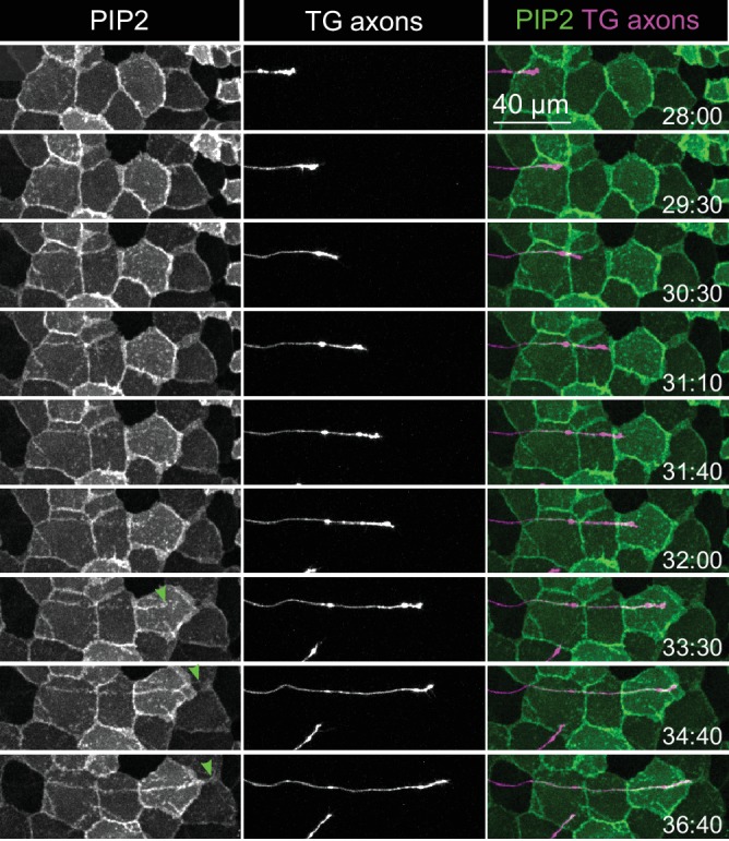
Figure 5—figure supplement 2. PIP2 microdomains form shortly after axons innervate the zebrafish epidermis.

Live-imaging of PIP2 in basal cells [TgBAC(tp63:GAL4FF); Tg(UAS:GFP-PH-PLC)] and trigeminal axons [Tg(isl1[ss]:LEXA-VP16,LEXAop:tdTomato)] during axon growth into the epidermis. Timestamp indicates HH:MM. Green arrowheads mark a growing sheath.

Acknowledgments
This image is the copyrighted work of the attributed author or publisher, and ZFIN has permission only to display this image to its users. Additional permissions should be obtained from the applicable author or publisher of the image. Full text @ Elife