
|
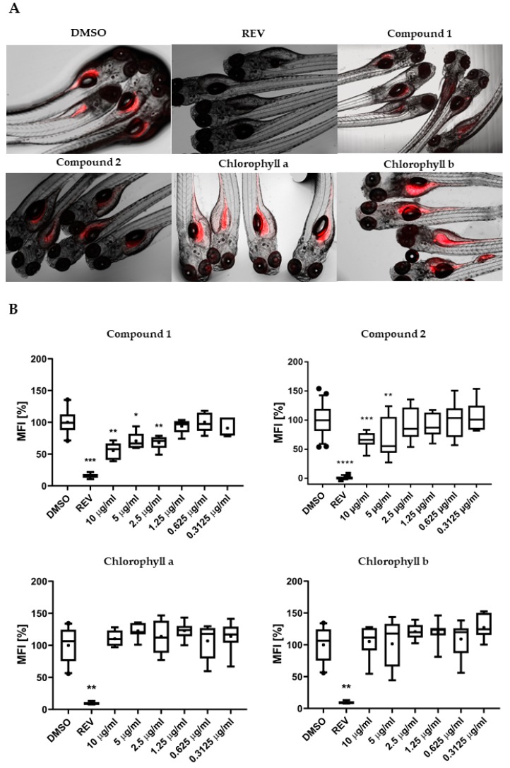
Figure 3
(A) Representation of the zebrafish Nile red fat metabolism assay. Strong fluorescence signal is present in zebrafish larvae from the solvent control around the yolk sac and stomach/intestine. Compounds 1 and 2 decreased the Nile red staining, in contrast to chlorophyll a and b. (B) Quantification of lipid-reducing activity in the zebrafish Nile red fat metabolism assay after exposure over 48 h. Solvent control was 0.1% dimethyl sulfoxide (DMSO) and positive control was 50 µM resveratrol (REV). Values are expressed as mean fluorescence intensity (MFI) relative to the DMSO group and are derived from six to eight individual larvae per treatment group. The data are represented as box-whisker plots from the fifth to 95th percentiles. Asterisks highlight significant altered fluorescence intensities that indicate changes of neutral lipid level (**** p < 0.0001; *** p < 0.001; ** p < 0.01; * p < 0.05).