IMAGE

Figure 1

ID
ZDB-IMAGE-190723-2526
Source
Figures for Amigo et al., 2018
Image
Figure Caption

Figure 1

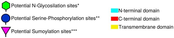
Domain structure and potential post-translational modification sites of human RPRM and RPRML proteins. Schematic representation shows the RPRM and RPRML putative N-glycosylation (green hexagons), serine-phosphorylation (blue circle) and sumoylation (purple triangle) sites. The N-terminal, transmembrane and C-terminal domains are represented by colored boxes (turquoise, yellow, and red, respectively). An * (asterisk) indicates positions which have a single, fully conserved residue. A: (colon) indicates conservation between groups of strongly similar properties—scoring >0.5 in the Gonnet PAM 250 matrix. A. (period) indicates conservation between groups of weakly similar properties—scoring 0.5 in the Gonnet PAM 250 matrix. Post-translational modification sites were predicted using *NetNGlyc (http://www.cbs.dtu.dk/services/NetNGlyc/), **NetPhos (http://www.cbs.dtu.dk/services/NetPhos/) and ***SumoPlot (http://www.abgent.com/sumoplot).

Acknowledgments
This image is the copyrighted work of the attributed author or publisher, and ZFIN has permission only to display this image to its users. Additional permissions should be obtained from the applicable author or publisher of the image. Full text @ Int. J. Mol. Sci.