IMAGE

Figure 1

ID
ZDB-IMAGE-190723-2239
Source
Figures for Wood et al., 2018
Image
Figure Caption

Figure 1

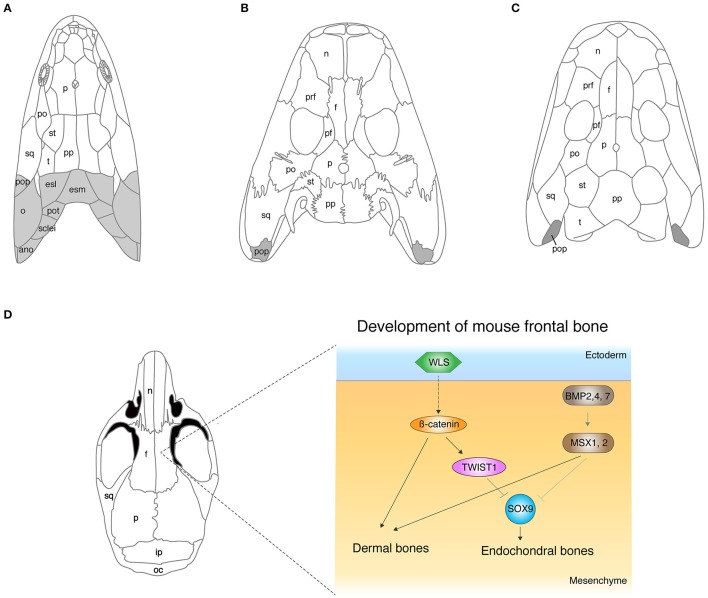
The evolution of cranial dermal bones and the developmental mechanisms. (A–C) The skull roof of Eusthenopteron(A), Acanthostega(B), Ichthyostega(C). (D) The developmental mechanisms of the frontal bone in mice. The molecules that initiate dermal differentiation from the ectoderm to the mesenchyme have not been identified yet, though the WNT signaling pathway is suggested to be involved (Goodnough et al., 2014). ano, anocleithrum; esl, lateral extrascapular; esm, medial extrascapular; f, frontal; ip, interparietal; n, nasal; o, opercular; oc, occipital; p, parietal; pf, postfrontal; po, postorbital; pop, preopercular; pot, post-temporal; pp, postparietal; prf, prefrontal; st, supratemporal; sq, squamosal; sclei, supracleithrum; t, tabular. Illustrations in (A–D) are redrawn with permissions from Clack (2012), Jarvik (1980), and White et al. (2016).

Acknowledgments
This image is the copyrighted work of the attributed author or publisher, and ZFIN has permission only to display this image to its users. Additional permissions should be obtained from the applicable author or publisher of the image. Full text @ Front Cell Dev Biol