
Image
|
Figure Caption
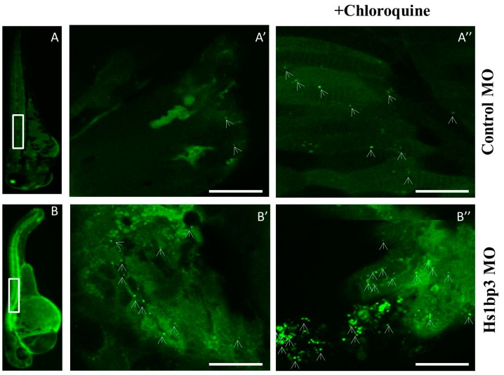
Figure 3
Confocal imaging of Tg(CMV:GFP-Lc3). Representative confocal images of GFP-Lc3 puncta (autophagosomes) in the trunk area of GFP-Lc3 transgenic zebrafish embryos injected with control morpholino or Hslbp3 translational-blocking morpholino and imaged at 2 days post fertilization (dpf) with or without pre-treatment with chloroquine (10 mM) for 6 h. Scale bars, 10 µM for the confocal images. Panel A, B shows the whole zebrafish larvae at 2 days post fertilization highlighting the trunk area chosen for confocal imaging; Panel A’, A’’, B’, B’’ shows respective confocal images.
Acknowledgments
This image is the copyrighted work of the attributed author or publisher, and
ZFIN has permission only to display this image to its users.
Additional permissions should be obtained from the applicable author or publisher of the image.
Full text @ Cells