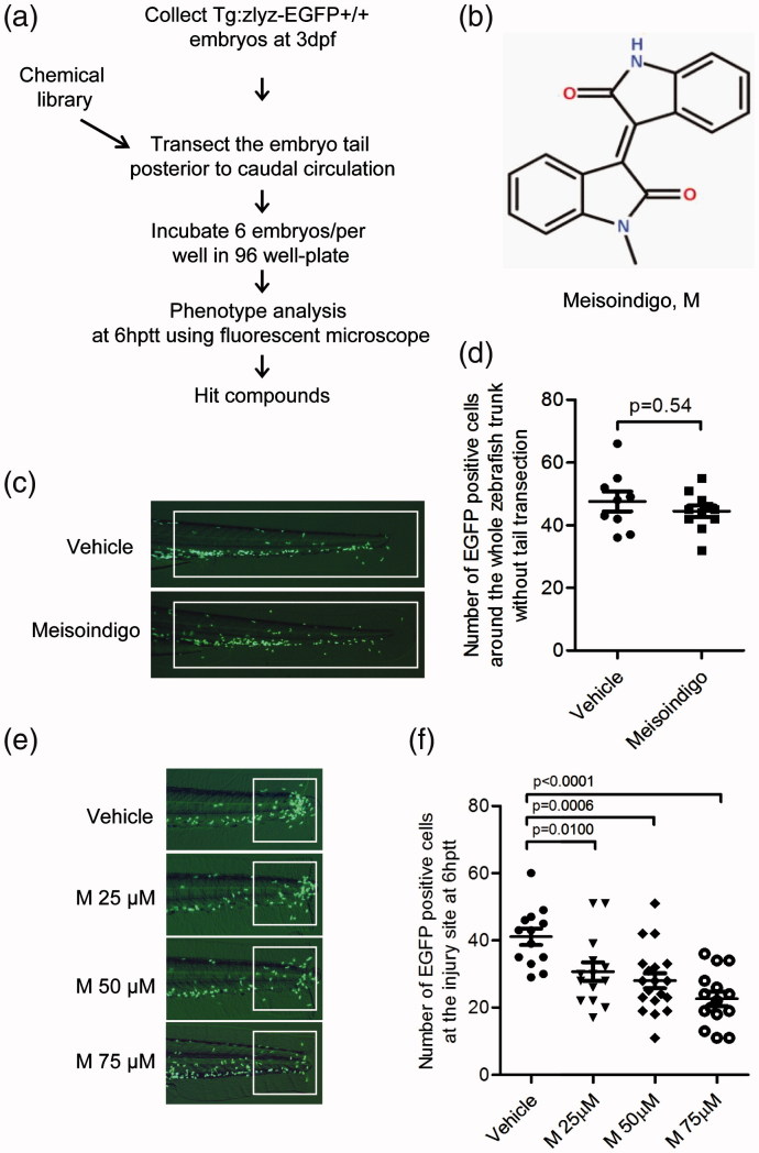
IMAGE

Figure 2.

ID
ZDB-IMAGE-190723-1877
Source
Figures for Ye et al., 2017
Image
Figure Caption

Figure 2.

Identification of meisoindigo that inhibits zebrafish leukocyte recruitment to the injury site. (a) Chemical screening strategy. (b) Chemical structure of meisoindigo. (c,d) Tg:zlyz-EGFP embryos (3 dpf) without tail transection were treated with vehicle(n = 9) or meisoindigo(M, 100 μM, n = 11) for 6 h. Leukocytes around the whole trunk of zebrafish embryo [white boxes in (c)] were quantitiatively analyzed. (e,f) Tg:zlyz-EGFP embryos (3 dpf) with tail transection were treated with vehicle (n = 13), 25 μM (n = 13), 50 μM (n = 18) or 75 μM (n = 14) meisoindigo, respectively. Leukocytes that migrated to the highly inflamed regions [white boxes in (e)] at 6 hptt were quantitatively analyzed (f). The p values were annotated as obtained from a two-tailed t test.

Acknowledgments
This image is the copyrighted work of the attributed author or publisher, and ZFIN has permission only to display this image to its users. Additional permissions should be obtained from the applicable author or publisher of the image. Full text @ Pharm Biol