IMAGE

Figure 4

ID
ZDB-IMAGE-190723-176
Source
Figures for Kawahara et al., 2016
Image
Figure Caption

Figure 4

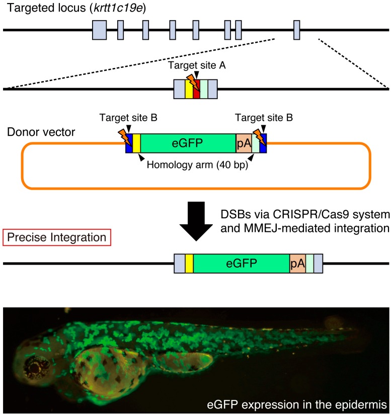
Precise site-specific integration of eGFP into the krtt1c19e locus. The donor vector consists of two bait sequences (blue box; target site B for Gbait-gRNA), a left homology arm (yellow box; 40 bp), and an eGFP (green box) and polyA signal (pA) (orange box) and right homology arm (light green box). The target site A for krtt1c19e-gRNA (red box) is located near the stop codon of the krtt1c19e gene. When the CRISPR/Cas9 system simultaneously cleaves the target sites A and B, the eGFP reporter between the microhomology arms is precisely integrated into the krtt1c19e locus. Broad eGFP expression was detected in the epidermis of the injected F0 embryo as described previously [44]. Gray boxes; exons in the krtt1c19e gene.

Acknowledgments
This image is the copyrighted work of the attributed author or publisher, and ZFIN has permission only to display this image to its users. Additional permissions should be obtained from the applicable author or publisher of the image. Full text @ Int. J. Mol. Sci.