
Image
|
Figure Caption
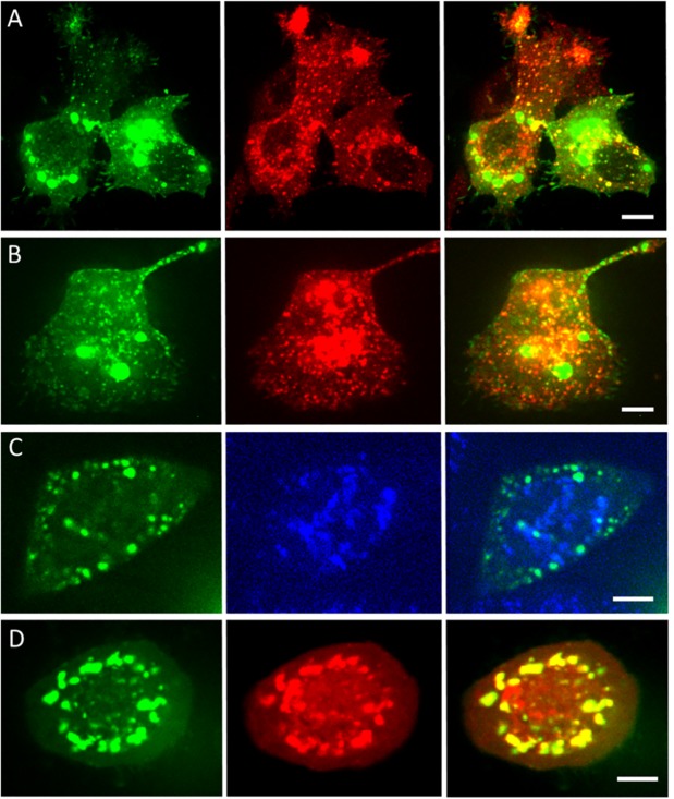
Fig 5
Two-channel imaging of turboGFP:MG53 (green fluorescence excited with 488-nm light, first column) with (second column) early endosomes, excited with 561-nm light (A), late endosomes, excited with 561-nm light (B), lysosomes, excited with 405-nm light (C), and inclusion bodies, excited with 561-nm light (D). Overlay images are shown in the third column. Scale bars, 10 μm.
Acknowledgments
This image is the copyrighted work of the attributed author or publisher, and
ZFIN has permission only to display this image to its users.
Additional permissions should be obtained from the applicable author or publisher of the image.
Full text @ PLoS One