
|
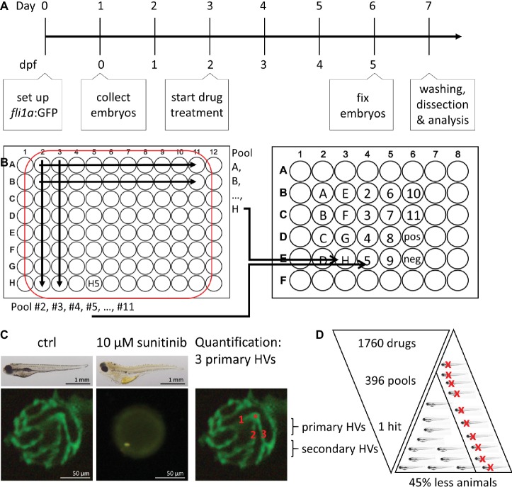
Figure 1
Method overview for orthogonal drug pooling. (A) Overview of drug treatment protocol. Tg(fli1a:eGFP) positive embryos were treated from 2–5 dpf and screened for intraocular vascular defects to assess the antiangiogenic potential of test chemicals. Sunitinib was used as positive antiangiogenic control and 1% DMSO as negative vehicle control. (B) In each 96 well plate, the 8 compounds of 10 columns and the 10 compounds of 8 rows were assembled in pools (left panel). This orthogonal pooling protocol reduced 80 individual compounds to 18 test pools. Every compound is represented in two pools (right panel). (C) The primary hyaloid vessel assay readout assesses the lenses dissected (lower left and middle box) from fixed larvae and quantification of the number of primary hyaloid vessels emerging from the optic disk (asterisk) on the back of the lens counted manually under a stereomicroscope (lower right box). (D) In total, 1,760 compounds were analyzed, combined in 396 pools, resulting in one confirmed hit using this method. This assay replaces animal use with immature larval forms and the orthogonal pooling reduces the number of immature larvae needed by 45%.