IMAGE

Figure 1

ID
ZDB-IMAGE-190723-1210
Source
Figures for Jagau et al., 2019
Image
Figure Caption

Figure 1

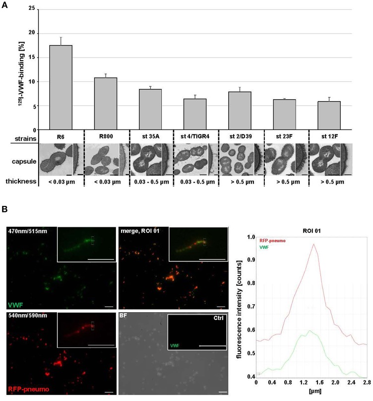
Streptococcus pneumoniae binds human plasma VWF. (A) Binding analyses with radioactive labeled VWF and seven S. pneumoniae strains: R6, R800 and the serotypes (st) 35A, 4 (TIGER4), 2 (D39), 23F and 12F. VWF binding was expressed as percentage of total radioactivity added. Amount of capsular polysaccharides (CPS) was categorized according to the CPS-diameter determined by electron microscopic visualization: low encapsulation <0.03μm, middle encapsulation in a range of 0.03–0.5μm and high encapsulation >0.5μm. Scale bars represent 0.5 μm in the overview figures and 0.1 μm in magnified sections. (B) Immunofluorescence visualization of VWF binding to RFP-expressing pneumococci with FITC-conjugated VWF-specific antibodies after embedding of VWF-incubated pneumococci in agarose. RFP-expressing bacteria appear in red at 540/590 nm and VWF-detection is visualized in green at 470/515 nm. VWF coated bacteria appear in yellow in the merged channel. The insets visualize a representative fluorescence signal at 3-fold magnification. Antibody controls revealed no unspecific VWF-signal detection on the surface of pneumococci (Ctrl). Scale bar represents 10 μm. Fluorescence intensity determination visualizes the VWF-bacterial colocalization within the region of interest (ROI 01).

Acknowledgments
This image is the copyrighted work of the attributed author or publisher, and ZFIN has permission only to display this image to its users. Additional permissions should be obtained from the applicable author or publisher of the image. Full text @ Front Microbiol