IMAGE

Figure 4

ID
ZDB-IMAGE-190723-1103
Source
Figures for Liao et al., 2018
Image
Figure Caption

Figure 4

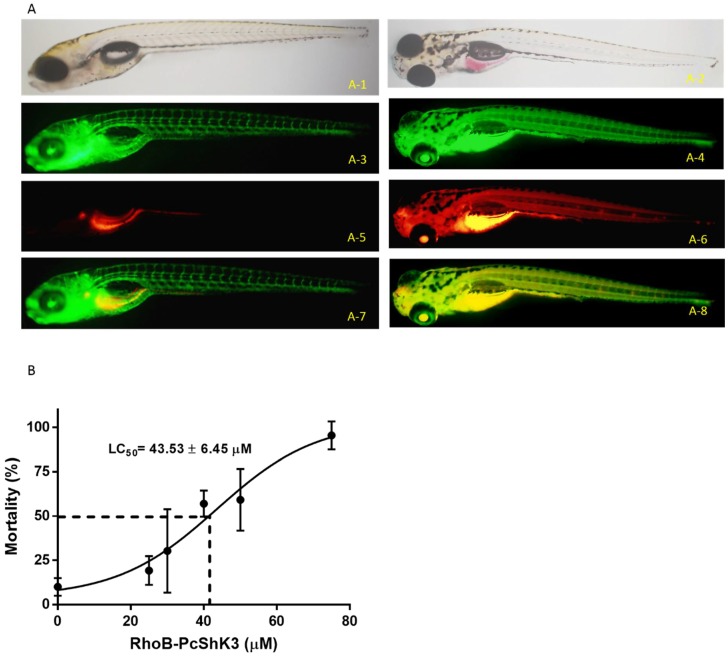
Mortality rate and distribution of P. caribaeorum ShK toxin-like peptide. (A) Fluorescent images of in vivo distribution of rhodamine B-conjugated peptide in Tg(fli1:EGFP)y1 zebrafish. Bright field pattern of zebrafish (6 dpf) treated with 20 μM of RhoB-PcShK3 (A-1) and free rhodamine B as negative control (A-2) for 24 h. Green fluorescence pattern of EGFP expression in transgenic Tg(fli1:EGFP)y1 zebrafish (B-3,B-4). Fluorescent images (A-5,A-6) from zebrafish (6 dpf) after incubation with rhodamine B-conjugated PcShK3 (20 μM) and free rhodamine B (20 μM) for 24 h. Merged images (A-7) of blood vessels (green) and rhodamine B conjugated PcShK3 (red). Merged fluorescent images (A-8) of blood vessels (green) and free rhodamine B (red); (B) Mortality rate of zebrafish larvae after exposure to peptide. The mortality rate reached 50% when zebrafish larvae was exposed to peptide (40 µM) for 8 h. The mortality rate reached 100% of zebrafish larvae exposed to peptide (75 µM) for 48 h. The LD50 is estimated to 43.53 ± 6.45 µM.

Acknowledgments
This image is the copyrighted work of the attributed author or publisher, and ZFIN has permission only to display this image to its users. Additional permissions should be obtained from the applicable author or publisher of the image. Full text @ Toxins (Basel)