
1
- ID
- ZDB-IMAGE-190723-1040
- Publication
- Greenhough et al., 2018 - Cancer cell adaptation to hypoxia involves a HIF-GPRC5A-YAP axis
- All Figures
- Figures for Greenhough et al., 2018
|
1
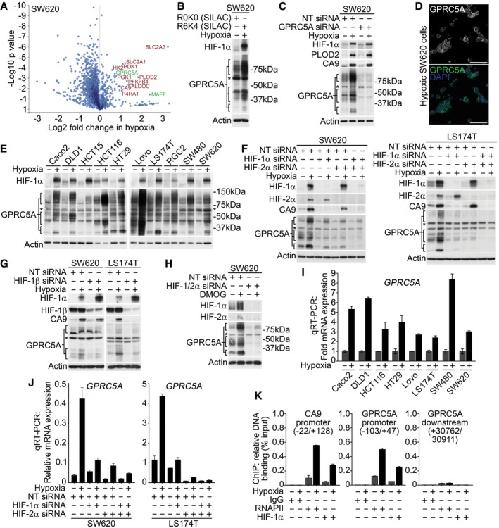
SILAC‐based proteomics data identify known (red) and novel (green) hypoxia‐induced proteins in SW620 cells. One‐sample Western blotting confirmed GPRC5A as a hypoxia‐induced protein in SILAC lysates. Validation of GPRC5A Western blot data using siRNA. *Non‐specific band of ˜60 kDa not depleted by GPRC5A siRNA. Confocal microscopy showing plasma membrane GPRC5A expression in hypoxic SW620 cells (scale bars: 75 μm). Western blotting showing GPRC5A upregulation by hypoxia in a panel of colorectal tumour cell lines. Basal & hypoxia‐induced GPRC5A protein expression was decreased by HIF‐1/2α depletion. Depletion of HIF‐1β decreased GPRC5A protein upregulation in hypoxia. Hypoxia mimetic DMOG induced HIF‐1/2α, CA9 and GPRC5A protein expression. Dual HIF‐1/2α depletion reduced GPRC5A induction by DMOG. qRT–PCR demonstrating that qRT–PCR demonstrating that HIF‐1/2α depletion decreased ChIP‐PCR analyses identify HIF‐1α binding to the
Data information: Asterisks (*) indicate non‐specific band. Level adjustments were made to images in Adobe Photoshop post‐acquisition for clarity (equal changes applied to the entire image). Representative examples of