
|
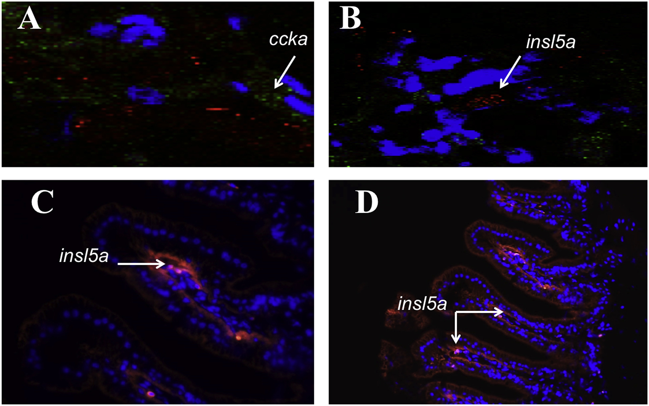
Fig. 5
In situ hybridization results in medaka. Cellular level of expression of ccka (positive control) (A) and insl5 (B) in enteroendocrine cells (EECS) of the gut following sectioning of frozen tissue in 5 μm thick slices using a cryostat microtome. Cells were incubated with a nuclear fluorescent stain (DAPI), and then hybridized with insl5a (red) and ccka(green) probes (see methods). Paraffin embedded sections were also used to assess the spatial localization of insl5aexpression (C, D). Paraffin blocks of medaka intestine were sectioned via microtome, and in situ hybridization performed using the insl5a antisense Stellaris probes and co-stained with a nuclear fluorescent stain (DAPI), revealing the location of insl5a probe in EECs.
Reprinted from Molecular and Cellular Endocrinology, 487, Alnafea, H., Vahkal, B., Zelmer, C.K., Yegorov, S., Bogerd, J., Good, S.V., Japanese medaka as a model for studying the relaxin family genes involved in neuroendocrine regulation: Insights from the expression of fish-specific rln3 and insl5 and rxfp3/4-type receptor paralogues, 2-11, Copyright (2019) with permission from Elsevier. Full text @ Mol. Cell. Endocrinol.