
|
Fig. 2
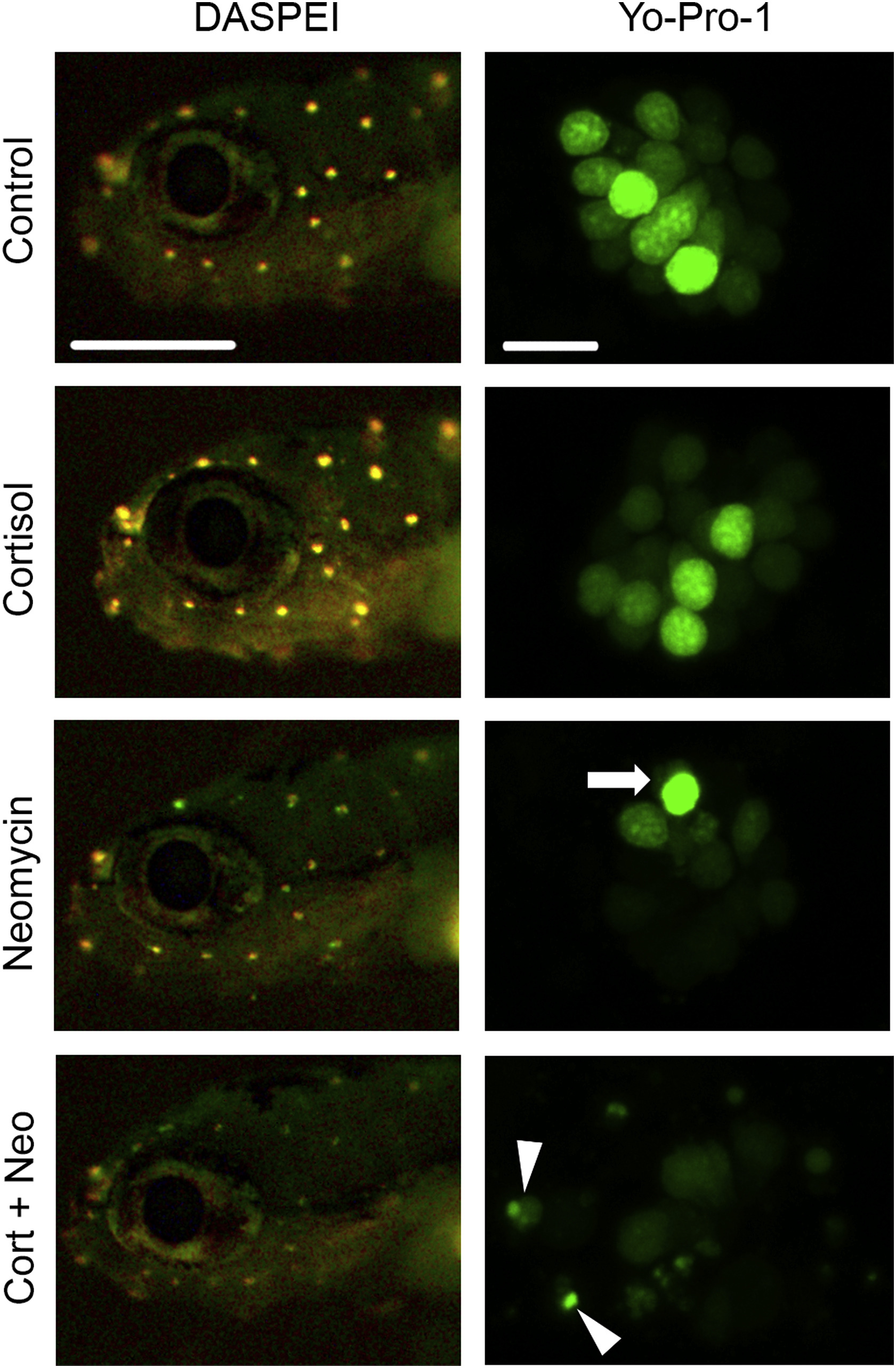
Cortisol increases neomycin-induced hair cell damage.Left column shows heads of fish labeled with the vital dye DASPEI. 50 μM neomycin treatment reduces DASPEI fluorescence, which is further reduced by 24-hr incubation with 10 μM cortisol. Scale bar in right column = 400 μm and applies to all DASPEI images. Right column shows higher magnification images of individual neuromasts (all of neuromast SO2, see Raible and Kruse, 2000) labeled with the nuclear-specific vital dye Yo-Pro-1. Incubation in 50 μM neomycin reduces the total number of hair cell nuclei and leads to formation of pyknotic nuclei (arrow). 24-hr incubation in 10 μM cortisol prior to neomycin exacerbates hair cell damage. Arrowheads indicate fragmented hair cell nuclei. Scale bar in left column = 10 μm and applies to all Yo-Pro-1 images.
Reprinted from Hearing Research, 377, Hayward, T., Young, A., Jiang, A., Crespi, E.J., Coffin, A.B., Glucococorticoid receptor activation exacerbates aminoglycoside-induced damage to the zebrafish lateral line, 12-23, Copyright (2019) with permission from Elsevier. Full text @ Hear. Res.