
Fig. 6
- ID
- ZDB-IMAGE-190708-12
- Publication
- Morales et al., 2019 - Peripheral Macrophages Promote Tissue Regeneration in Zebrafish by Fine-Tuning the Inflammatory Response
- All Figures
- Figures for Morales et al., 2019
|
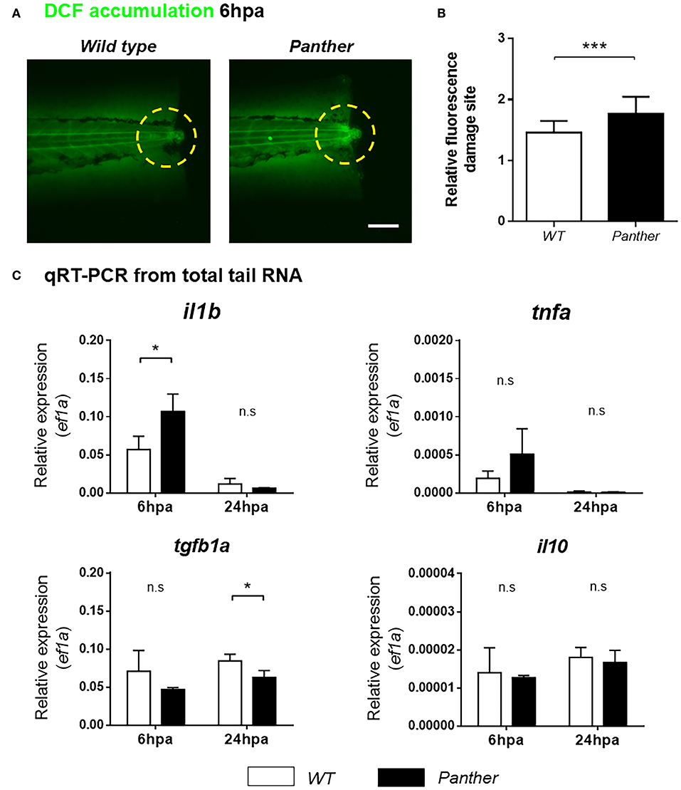
Fig. 6
Heightened il1b expression and ROS in the damage site of panther larvae after tail fin amputation. (A) The amount of ROS generated in the damage site (yellow dashed circle) was measured through the accumulation of the fluorescent 2′7′-dichlorofluorescein (DCF) sensor. Scale bar = 100 μm. (B) Relative fluorescence in the damage site ± SD of panther and wild type larvae. Twenty larvae per condition were analyzed. (C) Quantitative RT-PCR for il1b, tnfa, tgfb1a, and il10 from tails of panther and WT larvae at 6 and 24 hpa. The expression of ef1a was used as housekeeping for the 2−ΔCt calculation. A pool of ~20 larval tails was collected for RNA isolation, and the graphs show the mean ± SD of three independent experiments per condition. n.s not significant; *p < 0.05; ***p < 0.001.