
|
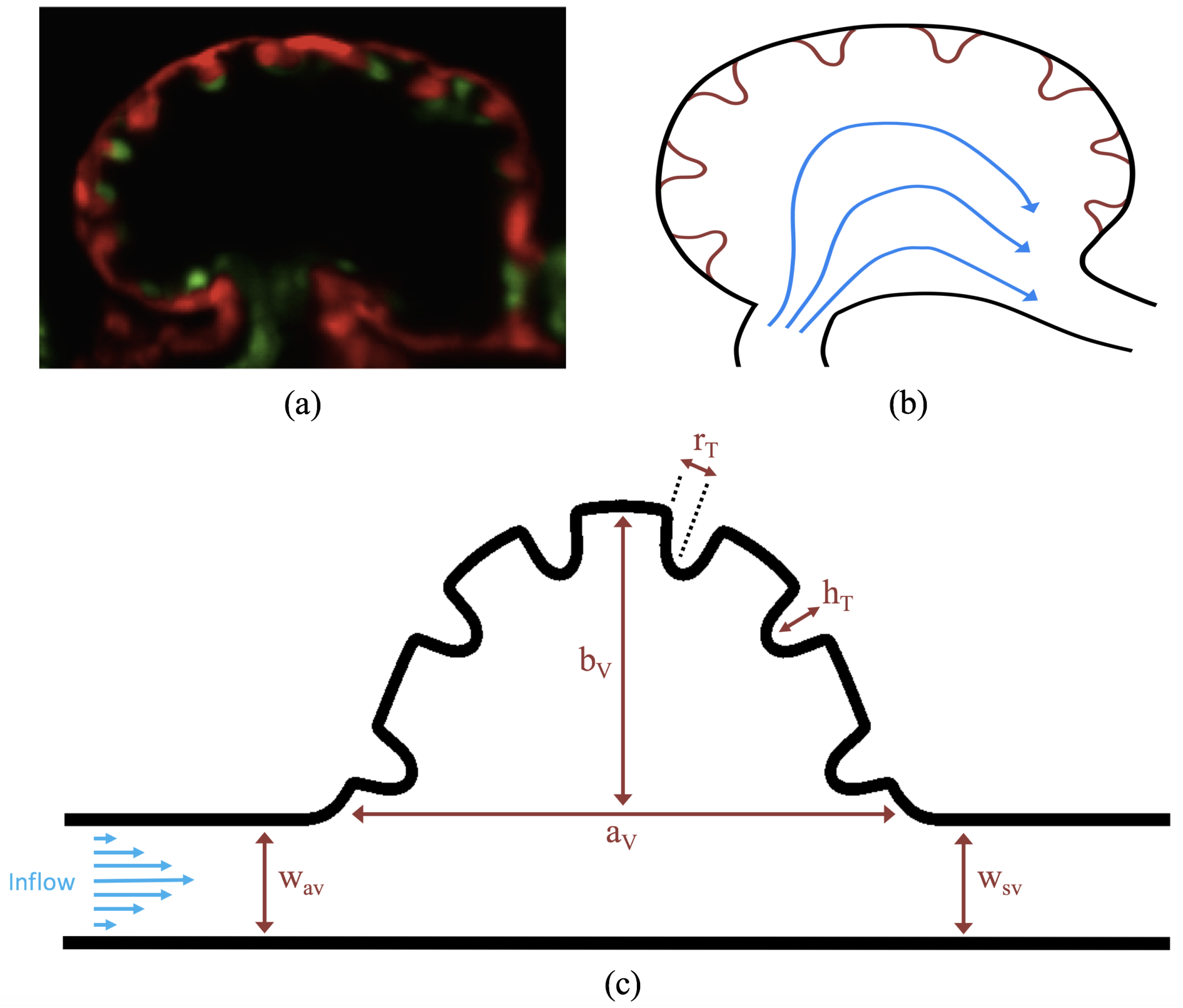
Fig. 3
(a) a microscopy image of an embryonic zebrafish’s trabeculated ventricle at 4 dpf from Liu et al. [21]. This snapshot was taken immediately before the systolic phase. The protrusions into the ventricle are trabeculae. The image was taken from Tg(cmlc2:dsRed)s879; Tg(flk1:mcherry)s843 embryos expressing fluorescent proteins that label the myocardium and endocardium, respectively [21]; (b) simplified diagram showing the basic idea behind our idealized geometry. Blood flows from the atrio-ventricular canal into the ventricle and then proceeds into the bulbus arteriosus; (c) further idealization of the computational model which is now flattened. The geometric parameters are as follows: aV and bV are the semi-major and semi-minor axis of the elliptical chamber, hT and rT are the height and radii of the trabeculae, and wAV and wSV are the widths of the atrioventricular (AV) canal and sinus venosus, respectively.