
|
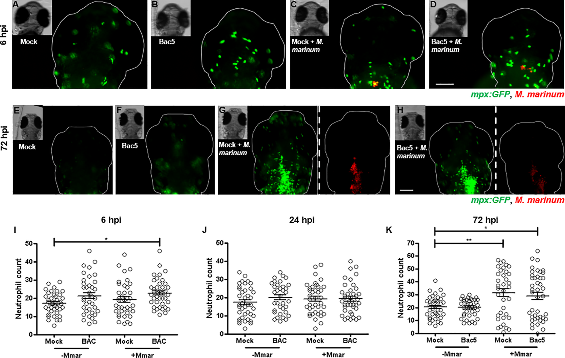
Fig. 4
Two days post-fertilisation casper Tg(mpx:GFP) zebrafish embryos were injected into their HBV with Mock, Bac5, Mock/M. marinum (Mmar) or Bac5/Mmar. 10 ng BAC was injected for both BAC treatment groups and approximately 250 CFU Mmar expressing DsRed2 for both infection groups. Z-stack images of the HBV region were acquired at 6 hpi, 24 hpi and 72 hpi. (A-D) Representative fluorescence overlay images of fish from a single experiment are shown at 6 hpi. Scale bar 100 μm. (E-H) Representative fluorescence images of fish from the same single experiment are shown at 72 hpi. Images of the bacterial fluorescence channel are shown only for infected zebrafish groups, separated by white dashed line from the neutrophil channel images. Scale bar 100 μm. (I-K) Neutrophils in the HBV region were quantified from fluorescence images of zebrafish embryos using Icy Spot Detector plugin. From L to R: 6 hpi, 24 hpi and 72 hpi. Data was also analysed using Icy FPC protocol with the same outcome (S5 Fig). Sample size (n) = 39, 37, 38, 43. Data pooled from three independent experiments is shown. Error bars represent S.E.M. One-way ANOVA with Bonferroni’s post-test. *p<0.05, **p<0.01, ***p<0.001.
https://doi.org/10.1371/journal.pone.0210508.g004