
IMAGE
Fig. S6
- ID
- ZDB-IMAGE-190626-39
- Publication
- Camargo-Sosa et al., 2019 - Endothelin receptor Aa regulates proliferation and differentiation of Erb-dependent pigment progenitors in zebrafish
- All Figures
- Figures for Camargo-Sosa et al., 2019
Image
|
Figure Caption
Fig. S6
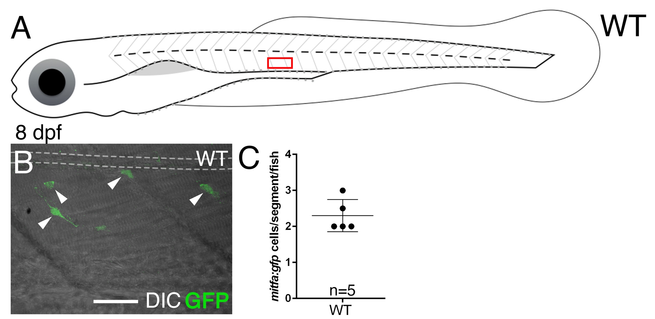
Expression of the Tg(mitfa:gfp) line in the ventral trunk of WT larvae.
(A) Scheme shows 8 dpf fish, with the red box indicating the area where mitfa:gfp positive cells in the ventral trunk were found. (B) GFP+ cells are readily found in the vicinity of the dorsal aorta throughout the posterior trunk and anterior tail at 8 dpf; superimposed DIC image shows these cells are not melanised. (C) Quantitation of GFP+ cells from a random posterior trunk segment in each of 5 fish, given as mean±s.d. = 2.3±0.44 (n = 5).
Acknowledgments
This image is the copyrighted work of the attributed author or publisher, and
ZFIN has permission only to display this image to its users.
Additional permissions should be obtained from the applicable author or publisher of the image.
Full text @ PLoS Genet.