
Fig. S1
- ID
- ZDB-IMAGE-190626-35
- Publication
- Camargo-Sosa et al., 2019 - Endothelin receptor Aa regulates proliferation and differentiation of Erb-dependent pigment progenitors in zebrafish
- All Figures
- Figures for Camargo-Sosa et al., 2019
|
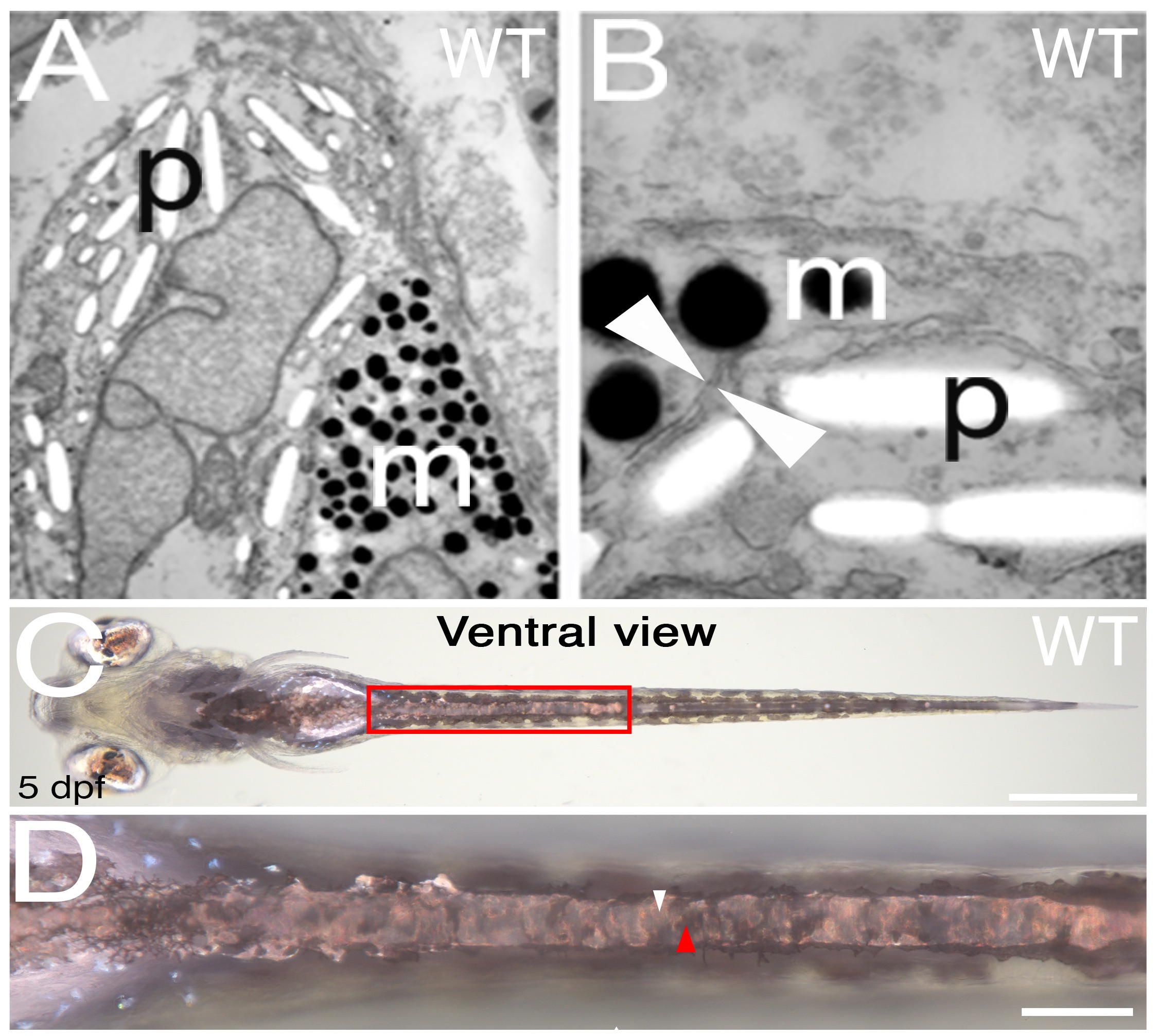
Fig. S1
Melanocytes and iridophores in the WT yolk sac stripe are consistently separated from each other by double membranes.
Transmission electron photomicrographs of melanocytes and iridophores in the WT yolk sac stripe ectopic pigment cells in pde mutants. A and B show two examples of melanosomes (m) and reflecting. platelets (p) separated by a double membrane (white arrowheads). Bright-field image of ventral view (C) and close up of the area in the red box (D) of WT fish, shows yolk sac stripe. Continuous layer of iridophores is indicated by white arrowhead in D, closely associated black melanocytes forming contiguous layer immediately dorsal to iridophores is indicatted by red arrowhead. Scale bars = 500 μm (C) and 100 μm (D).