
|
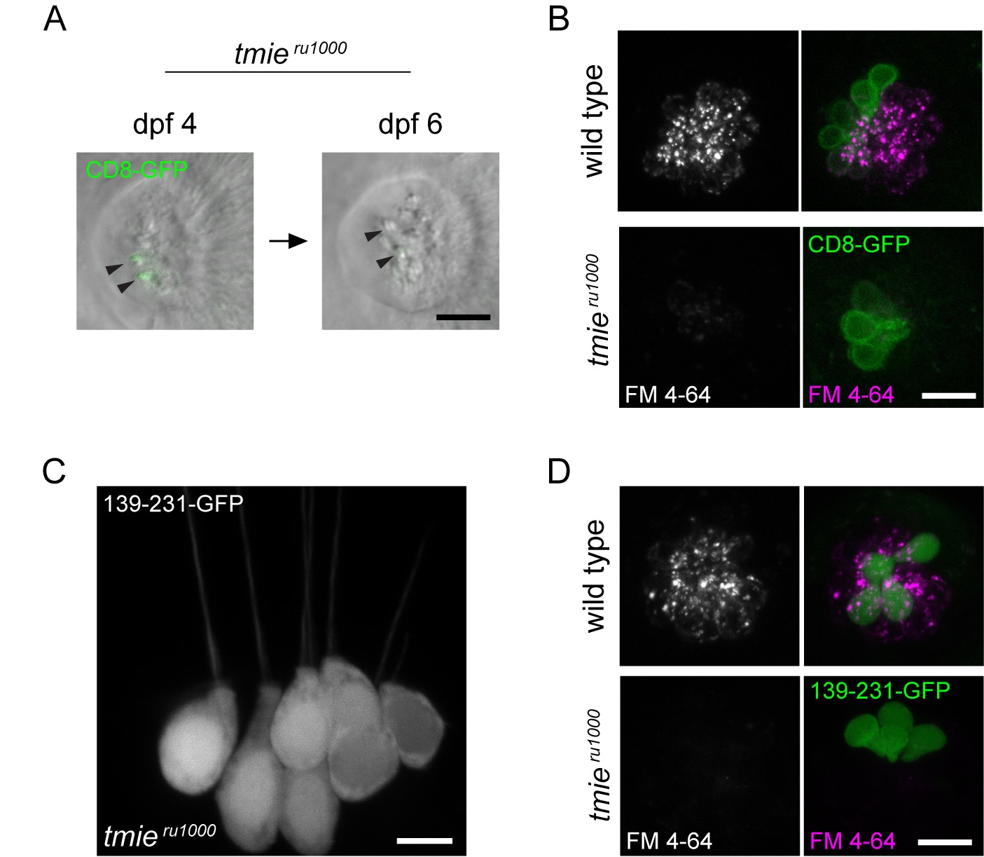
Fig. S4
Expression pattern and functional rescue by tmie constructs CD8 and 139–231.
All images were captured using confocal microscopy. (A) Stereocilia of a neuromast viewed from above. The same neuromast was imaged at 4 dpf and 6 dpf. In hair cells expressing CD8-GFP, signal was initially detected in immature bundles, but this expression was only detectable in soma by dpf 6 as the cells matured (n = 10 cells). (B) Maximum projection of neuromasts viewed from above; left panel shows only FM 4–64 while right panel adds CD8-GFP. No rescue of FM 4–64 labeling was observed in tmieru1000 hair cells expressing CD8-GFP (n = 40 cells). (C) Maximum projection of the posterior crista in a tmieru1000 larva with some hair cells expressing 139-231-GFP, which fills the cell (n = 43 cells). (D) Same as B except the transgene being expressed is 139-231-GFP. No rescue of FM 4–64 labeling was observed in tmieru1000hair cells expressing 139-231-GFP (n = 33 cells). Scale bars in A and C are 5μm, in B and D are 10μm.