
Fig. 3
- ID
- ZDB-IMAGE-190624-5
- Publication
- Huang et al., 2019 - Rab33a and Rab33ba mediate the outgrowth of forebrain commissural axons in the zebrafish brain
- All Figures
- Figures for Huang et al., 2019
|
Fig. 3
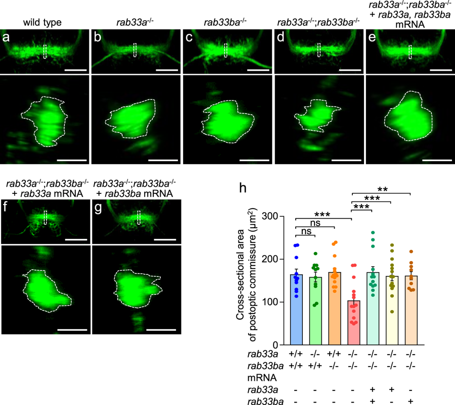
rab33a;rab33ba double mutants display a reduced cross-sectional area of the postoptic commissure. (a–g) Representative frontal views (upper panels) of the postoptic commissures of wild-type control (a), rab33a−/− single mutant (b), rab33ba−/− single mutant (c) and rab33a−/−;rab33ba−/− double mutant (d) embryos at 36 hpf. In (e), rab33a and rab33ba mRNAs were injected into a rab33a−/−;rab33ba−/− double mutant embryo for rescue analysis. In (f,g), rab33a mRNA (f) or rab33ba mRNA (g) were injected into a rab33a−/−;rab33ba−/− double mutant embryo for rescue analysis. Scale bars: 50 µm. The lower panels show the cross-sections at the dotted lines shown in the frontal views. Scale bars: 10 µm. (h) The cross-sectional area of the postoptic commissure obtained from the data analyses in (a–g; lower panels). Wild-type control (n = 11), rab33a−/− single mutant (n = 11), rab33ba−/− single mutant (n = 14) and rab33a−/−;rab33ba−/− double mutant (n = 14) embryos, and rab33a−/−;rab33ba−/− double mutant embryos with rab33a and rab33ba mRNAs (n = 13), rab33a mRNA (n = 14) and rab33ba mRNA (n = 10) were analyzed at 36 hpf. Data are mean ± SEM; ***P < 0.01; **P < 0.02; ns, not significant (one-way ANOVA with Tukey’s post hoc test).