
|
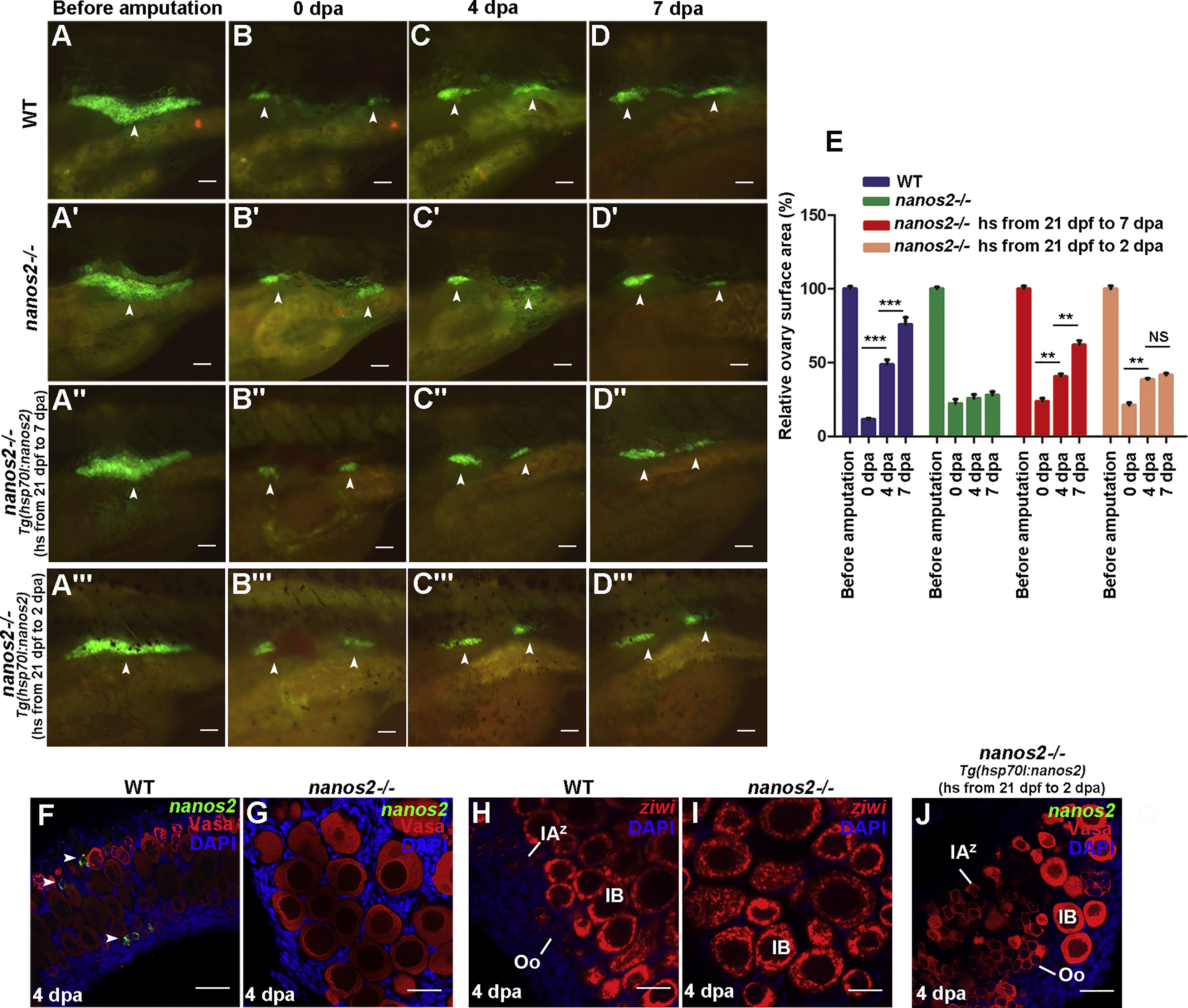
Fig. 4
The Ovary of nanos2 Mutant Fails to Regenerate
(A–D′) The ovaries of the wild-type juvenile under the Tg(vasa:DenNTR)cq41 transgenic background were imaged at before amputation/32 dpf (A), 0 (B), 4 (C), and 7 (D) dpa. The ovaries of nanos2 mutant were imaged at the same stages (A′–D′), showing little regeneration in contrast to the wild-type (A–D).
(A′–D′) The nanos2 mutants juvenile with Tg(vasa:DenNTRcq41; hsp70l:nanos2cq42) genetic background were heated shock from 21 dpf on and the ovaries were amputated at 32 dpf.
(A″–D″) Heat-shock (hs) is from 21 dpf to 7 dpa/39 dpf.
(A″′–D″′) Heat-shock is from 21 dpf to 2 dpa/34 dpf.
(E) Quantification of ovary surface area of wild-type and nanos2 mutant at 0, 4, and 7 dpa. (n = 5, mean ± SEM, ∗∗∗p < 0.001, ∗∗p < 0.01, NS represents no significance, two-tailed t test, error bars indicate SEM).
(F and G) Triple fluorescent labeling with FISH-nanos2, anti-Vasa antibodies, and DAPI staining in the ovaries of wild-type (F) and the nanos2 mutant (G) 4 dpa.
(H and I) Double labeling of FISH-ziwi and DAPI staining in the ovaries of wild-type (H) and nanos2mutant (I) at 4 dpa.
(J) Triple fluorescent labeling with nanos2 RNA probe, Vasa antibodies, and DAPI staining in the nanos2homozygous ovaries at 4 dpa, which were heat-shocked from 21 dpf to 2 dpa.
Scale bars: 500 μm (A–D″′), 50 μm (F–J).
(F–F″) Testis (arrowheads) that lack germ cells appears at 90 dpt, showing that the females treated with Mtz reverted to sterile males.
Oo, oogonia; IAz, zygotene-stage-IA; IA, stage IA oocytes; IB, stage IB oocyte; II, stage II oocyte; III, stage III oocyte; IV, stage IV oocyte; V, stage V oocyte. Scale bars: 50 μm (A), 1 mm (B–F′), 200 μm (B″–F″). See also Figure S2.