
Fig. 3
- ID
- ZDB-IMAGE-190620-13
- Publication
- Clément et al., 2018 - Cog4 is required for protrusion and extension of the epithelium in the developing semicircular canals
- All Figures
- Figures for Clément et al., 2018
|
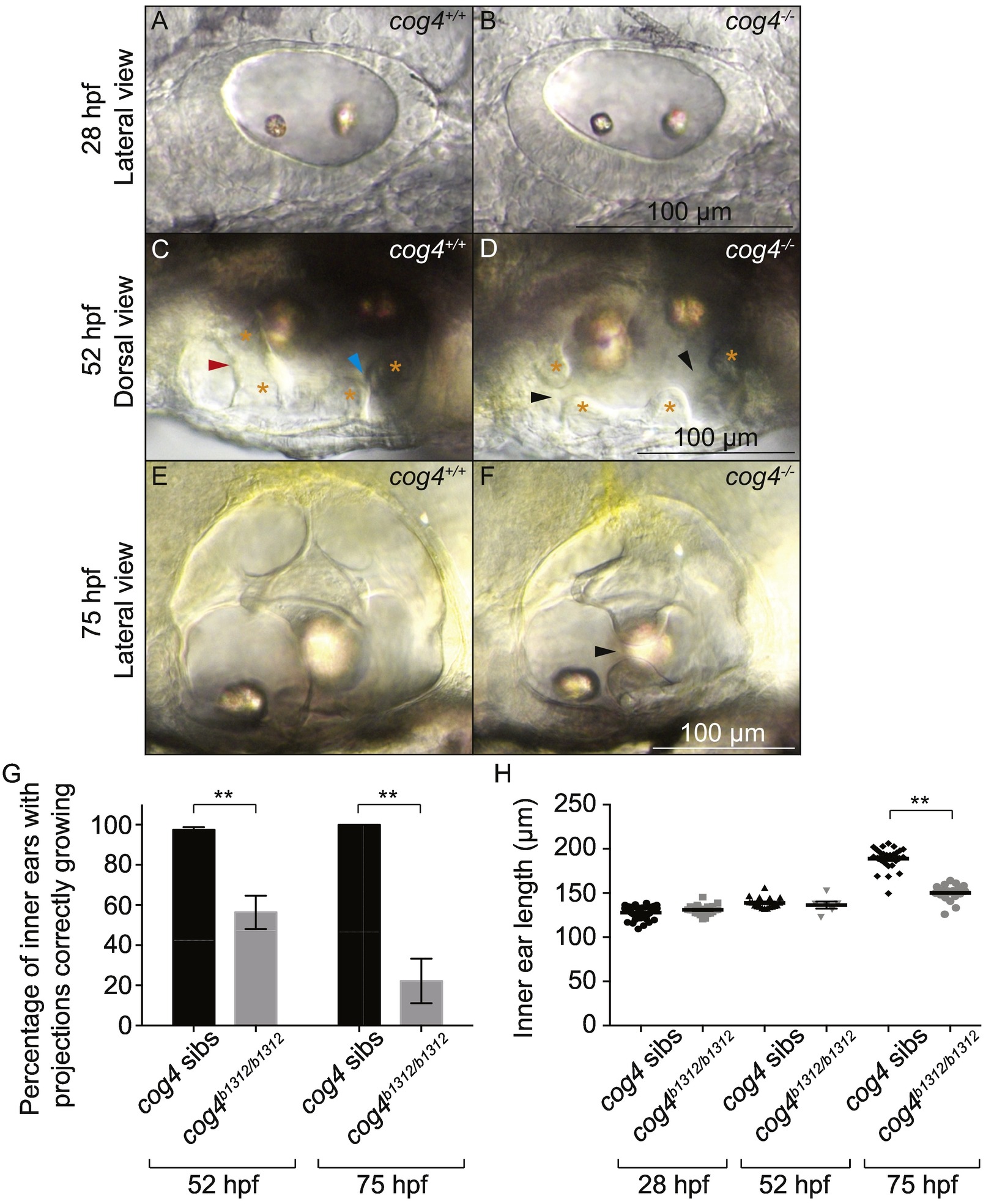
Fig. 3
Formation of the epithelial projections is delayed in cog4−/−mutants. Live images of the inner ear from cog4+/+ sibling (A, n = 49 embryos; C, n = 57 larvae; E, n = 26 larvae) and cog4−/− mutant larvae (B, n = 21 embryos; D, n = 22 larvae; F, n = 18 larvae) at 28 hpf (A, B), 52 hpf (C, D) and 75 hpf (E, F). Orange stars indicate the anterior and posterior projections and bulges (C, D). The red and blue arrowheads indicate the anterior and forming posterior pillar, respectively (C). Black arrowheads point to the gaps between the anterior and forming posterior projections and bulges (D, F). Anterior to the left (A–F). Dorsal to the top (A, B, E, F). Medial to the top (C, D). Percentage of inner ears with projections growing correctly at 52 hpf and 75 hpf, in cog4+/+ sibling (52 hpf, n = 114 inner ears; 75 hpf, n = 26 inner ears) and cog4−/− mutant larvae (52 hpf, n = 38 inner ears; 75 hpf, n = 18 inner ears) (G). Ear size at 28, 52 and 75 hpf in cog4+/+ sibling (28 hpf, n = 49 embryos; 52 hpf, n = 18 larvae; 75 hpf, n = 26 larvae) and cog4−/− mutant larvae (28 hpf, n = 21 embryos; 52 hpf, n = 6 larvae; 75 hpf, n = 18 larvae) (H). Size was measured along the antero-posterior axis of the inner ear. Data are represented as mean ± SEM. **p < 0.01.
Reprinted from Mechanisms of Development, 155, Clément, A., Blanco-Sánchez, B., Peirce, J.L., Westerfield, M., Cog4 is required for protrusion and extension of the epithelium in the developing semicircular canals, 1-7, Copyright (2018) with permission from Elsevier. Full text @ Mech. Dev.