
Fig. 2
- ID
- ZDB-IMAGE-190530-32
- Antibodies
- Publication
- Chen et al., 2019 - Degradation of endogenous proteins and generation of a null-like phenotype in zebrafish using Trim-Away technology
- All Figures
- Figures for Chen et al., 2019
|
Fig. 2
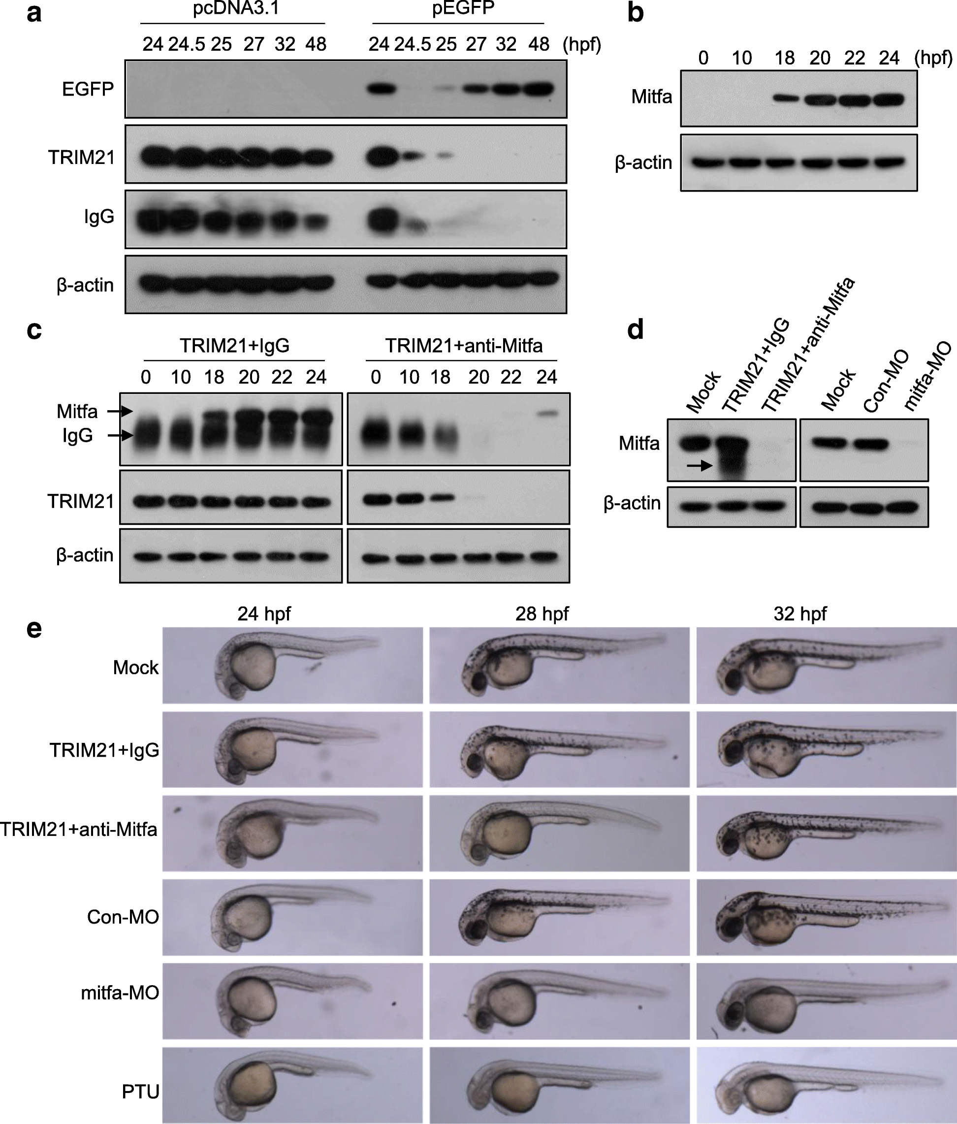
Degradation of Mitfa occurs 18 h after injection of TRIM21 and anti-Mitfa antibody. aEGFP expression plasmid or control pcDNA3.1 plasmid was injected into one-cell embryos. TRIM21 and anti-EGFP antibody were then co-injected into EGFP-expressing embryos or pcDNA3.1 plasmid-injected embryos at 24 hpf. The levels of EGFP, anti-EGFP antibody, and TRIM21 were analyzed by western blotting at different time points. bMitfa expression in zebrafish embryos was determined at different time points by western blotting. c One-cell embryos were co-injected with TRIM21 and an anti-Mitfa antibody or nonspecific IgG and analyzed for the levels of Mitfa, TRIM21, and IgG heavy chain at different time points by western blotting. d, e One-cell zebrafish embryos were injected with mitfa morpholinos (mitfa-MO) or control morpholinos (Con-MO) or co-injected with TRIM21/anti-Mitfa antibody or TRIM21/nonspecific IgG. Mitfa knockdown was confirmed at 22 hpf by western blotting; the arrow indicates IgG heavy chain (d). Lateral view of the embryos at different time points; embryos treated with N-phenylthiourea (PTU) were used as positive controls (e)