
IMAGE
Fig. S9
- ID
- ZDB-IMAGE-190530-18
- Publication
- Konjikusic et al., 2018 - Mutations in Kinesin family member 6 reveal specific role in ependymal cell ciliogenesis and human neurological development
- All Figures
- Figures for Konjikusic et al., 2018
Image
|
Figure Caption
Fig. S9
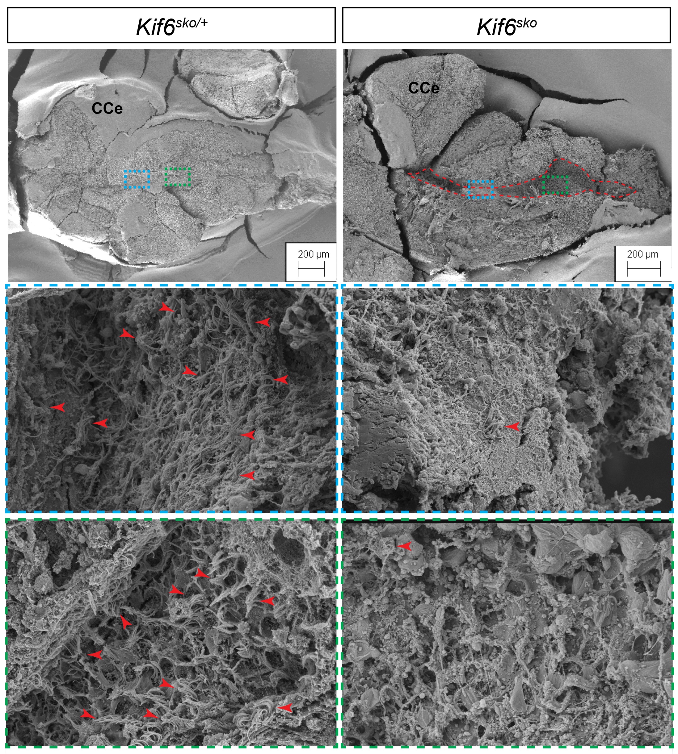
SEM of ventricle in kif6sko mutant zebrafish display dilation of the ventricular system and loss of ependymal cell cilia.
Scanning Electron Microscopy of zebrafish brain shows dilation of rhombencephalic (blue box) and telencephalic (green box) ventricles (red dotted line) indicative of hydrocephaly. Higher magnification images reveal loss of ependymal cell cilia tufts (red arrowheads) in kif6 zebrafish mutants when compared with heterozygous counterparts (red arrowheads). Scale bars 20μM and 200μM. (CCe: Cerebellum)
Figure Data
Acknowledgments
This image is the copyrighted work of the attributed author or publisher, and
ZFIN has permission only to display this image to its users.
Additional permissions should be obtained from the applicable author or publisher of the image.
Full text @ PLoS Genet.