
Fig. 5
- ID
- ZDB-IMAGE-190426-5
- Publication
- Anbalagan et al., 2018 - Pituicyte Cues Regulate the Development of Permeable Neuro-Vascular Interfaces
- All Figures
- Figures for Anbalagan et al., 2018
|
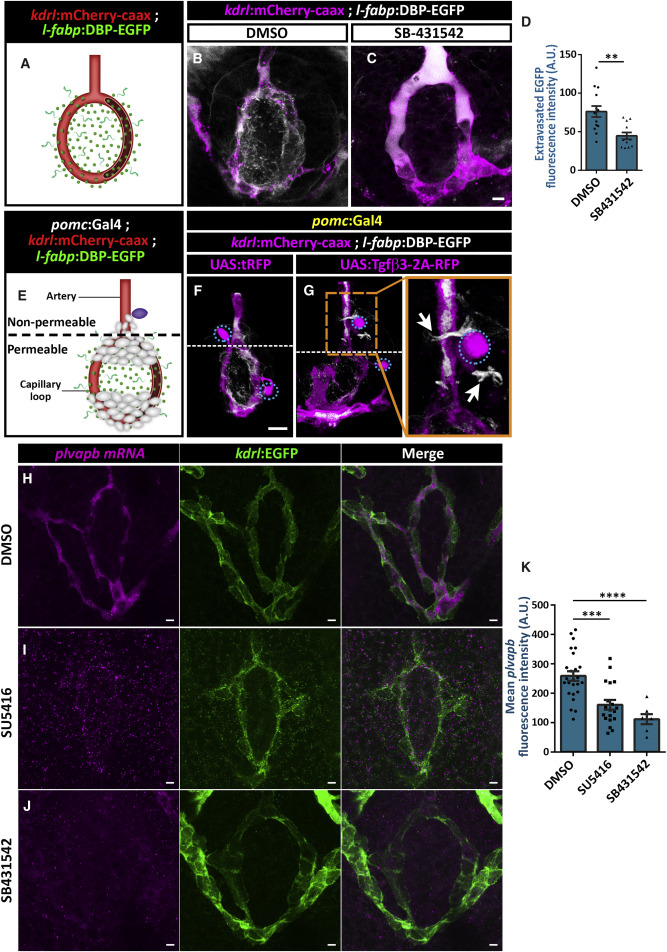
Fig. 5 Tgfβ and Vegf Signaling Increases Vascular Permeability and plvapb Expression (A–D) Measurement of hypophyseal permeability using the transgenic biosensor Tg(l-fabp:DBP-EGFP;kdrl:mCherry-caax) (A) was performed as described in Figure 4. Larvae were treated with either DMSO (B, n = 14) or TGF-β type I receptor inhibitor, SB431542 (C, n = 12), between 4 and 5 dpf, and extravasation of DBP-EGFP protein was quantified (D, ∗∗p < 0.01; Mann-Whitney test). A.U., arbitrary units. Scale bar: 5 μm. (E–G) The effect of Tgfβ3 on permeability was determined using the triple transgenic system Tg(l-fabp:DBP-EGFP;kdrl:mCherry-caax;pomc:Gal4) (E). The pomc:Gal4 driver was used to generate local hypophyseal clones overexpressing either UAS:tRFP (F, n = 10) or UAS:tgfβ3-2A-tRFP (G, n = 22), as described in Figure 4. Dashed lines mark the border between the permeable hypophyseal capillary and the non-permeable hypophyseal artery. Mosaic expression of Tgfβ3 (G, dotted cell) near the non-permeable artery led to extravasation of DBP-EGFP (inset; white arrows), whereas tRFP expression did not induce that effect (F). Correlation between the co-occurrence of artery extravasation and Tgfβ3 clones was determined by calculating the phi coefficient of association, followed by Fisher’s exact test of association (n = 8/22; p < 0.05) Scale bar: 10 μm. (H–K) Measurement of in situ plvapb expression upon Vegf or Tgf signaling perturbation. Confocal z stack images showing fluorescent in situ hybridization (FISH) of transgenic larvae using probes directed against mRNAs of plvapb. Larvae were treated with either DMSO (H, n = 24), SU5416 (I, n = 19) or SB-431542 (J, n = 6) between 4 and 5 dpf, and plvapb fluorescent intensity was quantified (K, ∗∗∗∗p < 0.0001; ∗∗∗p < 0.001; one-way ANOVA). A.U., arbitrary units. Scale bar: 5 μm. Data are presented as mean ± SEM (D and K). See related Figures S4 and S5.
Reprinted from Developmental Cell, 47(6), Anbalagan, S., Gordon, L., Blechman, J., Matsuoka, R.L., Rajamannar, P., Wircer, E., Biran, J., Reuveny, A., Leshkowitz, D., Stainier, D.Y.R., Levkowitz, G., Pituicyte Cues Regulate the Development of Permeable Neuro-Vascular Interfaces, 711-726.e5, Copyright (2018) with permission from Elsevier. Full text @ Dev. Cell