
Fig. 4
- ID
- ZDB-IMAGE-190426-4
- Publication
- Anbalagan et al., 2018 - Pituicyte Cues Regulate the Development of Permeable Neuro-Vascular Interfaces
- All Figures
- Figures for Anbalagan et al., 2018
|
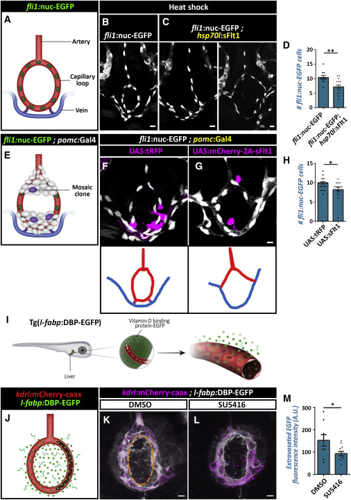
Fig. 4 Vegf Signaling Regulates Hypophyseal Capillary Morphogenesis and Permeability (A–D) Morphological analysis and cell count of the hypophyseal artery and capillary loop and vein using the Tg(fli1:nuc-EGFP) line, which labels endothelial cell nuclei (A). The transgenic line Tg(hsp70l:sFlt1) was used to inhibit Vegf/Vegfr2 signaling temporally. Control (B, n = 10) and Tg(hsp70l:sFlt1) larvae (C, n = 15) were subjected to multiple 28οC to 37οC temperature shifts between 3 dpf and 5 dpf. The capillary loop cell number (D) was significantly higher in Tg(hsp70l:sflt1) larvae (∗∗p < 0.01; Student’s t test). Correlation between the co-occurrence of abnormal loops and temporally induced sFlt1 expression was determined by calculating the phi coefficient of association, followed by Fisher’s exact test (n = 6/15; p < 0.03). Scale bars: 10 μm. (E–H) Hypophyseal clones (E) overexpressing control tRFP (F) or the soluble antagonist sFlt1 (E and G). Double transgenic Tg(pomc:Gal4;fli1:nuc-EGFP) embryos were injected with transposon-based transgenic expression vectors containing either control UAS:tRFP (n = 19) or UAS:mCherry-2A-sFlt1 (n = 15), allowing for mosaic gene expression in discrete hypophyseal cells (magenta). Cell numbers in the capillary loop (H) were significantly higher in the mutant (∗p < 0.05; Mann-Whitney test). The schemata shown at the bottom of panels depict the normal (F) and abnormal (G) connectivity of the hypophyseal capillary loop in 5 dpf larvae with mosaic expression. Correlation between the co-occurrence of the abnormal loop and sFlt1 clones was determined by calculating the phi coefficient of association, followed by Fisher’s exact test (n = 5/15; p < 0.05). Scale bars: 5 μm. (I–M) Assessment of the effect of Vegfr inhibitor, SU5416, on permeability in transgenic reporter Tg(l-fabp:DBP-EGFP;kdrl:mCherry-caax) larvae. D-vitamin binding protein (DBP) expressed in hepatocytes (l-fabp promoter) served as an endogenous permeability biosensor (I). Extravasation of the DBP-EGFP protein from the permeable neurohypophyseal vascular cells (J) was measured in the area inside the hypophyseal capillary loop (dashed line in K). Larvae were treated with DMSO (K, n = 9) or SU5416 at 5 μM (L, n = 13) between 4 and 5 dpf. Treatment with SU5416 led to reduced extravasation of DBP-EGFP from the hypophyseal loop (M, ∗p < 0.05; Mann-Whitney test; for each treatment). A.U., arbitrary units. Scale bar: 5 μm. Data are presented as mean ± SEM (D, H, and M). See related Figures S4 and S5.
Reprinted from Developmental Cell, 47(6), Anbalagan, S., Gordon, L., Blechman, J., Matsuoka, R.L., Rajamannar, P., Wircer, E., Biran, J., Reuveny, A., Leshkowitz, D., Stainier, D.Y.R., Levkowitz, G., Pituicyte Cues Regulate the Development of Permeable Neuro-Vascular Interfaces, 711-726.e5, Copyright (2018) with permission from Elsevier. Full text @ Dev. Cell