
Fig. 2
- ID
- ZDB-IMAGE-190426-2
- Publication
- Anbalagan et al., 2018 - Pituicyte Cues Regulate the Development of Permeable Neuro-Vascular Interfaces
- All Figures
- Figures for Anbalagan et al., 2018
|
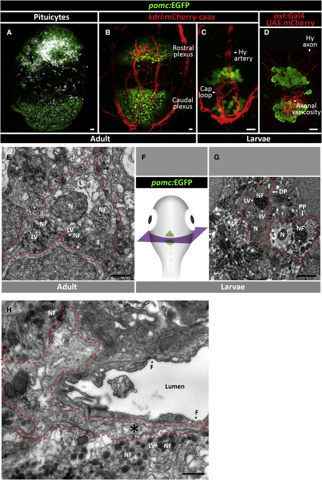
Fig. 2 Tissue Organization and Ultrastructure of Adult and Larval Pituicytes (A and B) Confocal maximal intensity image of a 3-month-old zebrafish hypophysis showing the spatial distribution of β-Ala-Lys-Nɛ-AMCA-labeled pituicytes (A), which intermingle with, but do not co-localize with adenohypophyseal proopiomelanocortin-(POMC) positive cells marked by the Tg(pomc:EGFP) transgenic reporter (A and B). The two main clusters of POMC+ cells that abut the pituicytes are located within the rostral and caudal hypophyseal Tg(kdrl:mCherry-caax)-labeled capillary plexus in the adult. Scale bars: 10 μM. (C and D) The spatial organization of the two anterior and posterior POMC-positive clusters in 9 dpf larva, where they abut the hypophyseal capillary loop and artery (C) and projecting neurohypophyseal axons (D), marked by Tg(oxt:EGFP). Scale bars: 10 μM. (E–H) Transmitted electron microscope (TEM) imaging of 3-month-old adult (E and H) and larval (9 dpf) (G) pituicytes. Correlative confocal imaging of hypophyseal POMC+ cells was used as an anatomical landmark to localize the position of adult and larval pituicytes prior to tissue sectioning (F) and TEM imaging. Representative pituicytes are delimited by red dashed lines. Note the pituicyte-specific cellular features such as a prominent nucleus [N], lipid droplets [L], and engulfment of nerve fibers [NF] that are filled with large electron-dense core vesicles [LV] and pituicyte projection [∗] separating nerve fibers from fenestrated capillaries [F]. DP, dense pituicyte; PP, pale pituicyte. Scale bars: 1 μm.
Reprinted from Developmental Cell, 47(6), Anbalagan, S., Gordon, L., Blechman, J., Matsuoka, R.L., Rajamannar, P., Wircer, E., Biran, J., Reuveny, A., Leshkowitz, D., Stainier, D.Y.R., Levkowitz, G., Pituicyte Cues Regulate the Development of Permeable Neuro-Vascular Interfaces, 711-726.e5, Copyright (2018) with permission from Elsevier. Full text @ Dev. Cell