
Fig. 1
- ID
- ZDB-IMAGE-190426-1
- Publication
- Anbalagan et al., 2018 - Pituicyte Cues Regulate the Development of Permeable Neuro-Vascular Interfaces
- All Figures
- Figures for Anbalagan et al., 2018
|
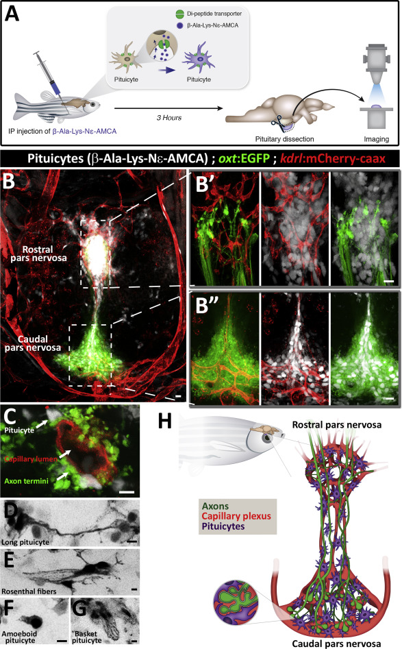
Fig. 1 Pituicytes, Axons, and Blood Capillaries Form Multiple Neurovascular Interfaces (A) Scheme describing functional labeling of pituicytes in vivo. Injection of the fluorescent aminomethylcoumarin-(AMCA) conjugated diamino acid peptide β-Ala-Lys-Nɛ intraperitoneally (IP) into adult zebrafish leads to active uptake of β-Ala-Lys-Nɛ-AMCA by pituicytes. 3 hr following injection, the hypophysis was dissected and imaged. (B) Whole-mount imaging of hypophysis dissected from 3-month-old transgenic reporter zebrafish allowing simultaneous visualization of neurohypophyseal axons, labeled by Tg(oxt:EGFP), and blood capillaries, labeled by Tg(kdrl:mCherry-caax), shows β-Ala-Lys-Nɛ-AMCA-labeled pituicytes. Scale bar, 5 μm. The axo-glial-vasal interface is seen in both the rostral (Bʹ) and caudal (Bʹʹ) pars nervosa of the neurohypophysis. Scale bar: 10 μm. (C) High magnification of a neurovascular unit showing pituicytes and neurohypophyseal axon termini interfacing with a single blood capillary. Scale bar: 5 μm. (D–G) Inverted gray scale high-magnification images of morphologically distinct pituicyte subtypes. Four major subtypes were identified: long processes connecting multiple pituicytes (D), pituicytes forming Rosenthal fibers (E), amoeboid pituicytes (F), and basket pituicytes (G). Scale bar: 5 μm. (H) Schematic three-dimensional model of the adult zebrafish neurohypophysis. Hypophyseal axons (green), blood capillaries plexus (red), and pituicytes (magenta) are the main components of the neurohypophysis. Axo-glial-vasal contacts are mainly present in the rostral and caudal pars nervosa. Inset: enlarged scheme of an axo-glio-vasal neurovascular unit depicting engulfment of axon swellings by pituicytes that also contact the perivascular space of blood capillaries.
Reprinted from Developmental Cell, 47(6), Anbalagan, S., Gordon, L., Blechman, J., Matsuoka, R.L., Rajamannar, P., Wircer, E., Biran, J., Reuveny, A., Leshkowitz, D., Stainier, D.Y.R., Levkowitz, G., Pituicyte Cues Regulate the Development of Permeable Neuro-Vascular Interfaces, 711-726.e5, Copyright (2018) with permission from Elsevier. Full text @ Dev. Cell