
|
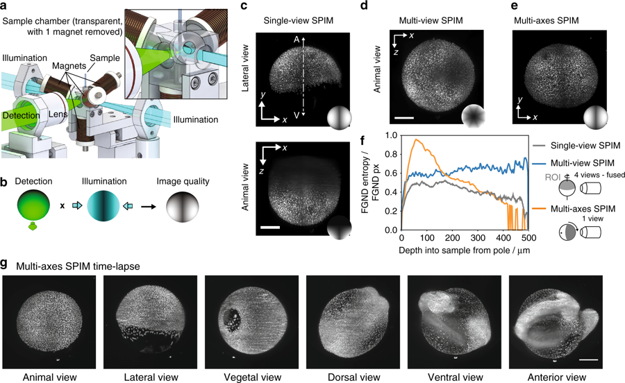
Fig. 4 Non-contact 3D orientation technique adapted to a SPIM system. a Schematic of the SPIM setup with the tetrahedral electromagnets arranged around the sample tube. The magnets were held by the sample chamber and the sample was illuminated by a light sheet through two windows. Fluorescence was detected through a third window by a detection objective. The sample chamber and the detection objective were motorized to move the sample through the light sheet and to correct for the different path lengths in air and water. b Schematic representation of the obtained image quality in SPIM imaging, which is influenced by the sample orientation relative to the detection and illumination objectives. c Maximum intensity projection of a single-view stack from a histone labeled zebrafish embryo. Scale bar, 200 μm. Inset: schematic representation of the image quality obtained by a single view. d Maximum intensity projection obtained by multi-view SPIM imaging of four views (0°, 45°, 180°, and 225°). The fusion of the four views shows a homogenous resolution along the equator but a decreasing resolution towards the cap (rotational axis). Scale bar, 200 μm. Inset: schematic representation of the image quality obtained by multi-view fusion of four views. e Maximum intensity projection of a single-view stack obtained with the multi-axis SPIM technique. The view on the animal pole shows the improved image quality of the animal pole (ROI) obtained by orienting the animal pole towards the detection objective by the multi-axis sample orientation technique. Scale bar, 200 μm. Inset: schematic representation of the image quality obtained with the multi-axis SPIM technique. f Local entropy as a measure of the image quality of the single-view, multi-view, and multi-axis data along the animal-vegetal axis. g Stills from a time-lapse of a developing, histone labeled zebrafish taken in different orientations to watch key events in the respective optimal orientation (Supplementary Movie 6). Scale bar, 200 μm