
Image
|
Figure Caption
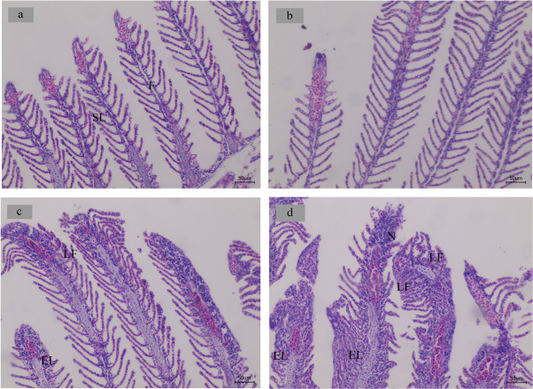
Fig. 5 Light micrographs of gill morphology of adult zebrafish exposed to maduramicin for 14 days (100 ×). (a) A control group exhibited normal structure, including a gill arch formed by filaments (F) and lamellae (SL); (b) gill exposed to 0.1 mg/L maduramicin exhibited normal histology; (c) gill exposed to 0.5 mg/L maduramicin exhibited slight epithelial lifting (EL) and lamellar fusion (LF); (d) gill exposed to 2.5 mg/L maduramicin exhibited serious epithelial lifting (EL), lamellar fusion (LF) and necrosis (N).
Acknowledgments
This image is the copyrighted work of the attributed author or publisher, and
ZFIN has permission only to display this image to its users.
Additional permissions should be obtained from the applicable author or publisher of the image.
Full text @ Ecotoxicol. Environ. Saf.