
Image
|
Figure Caption
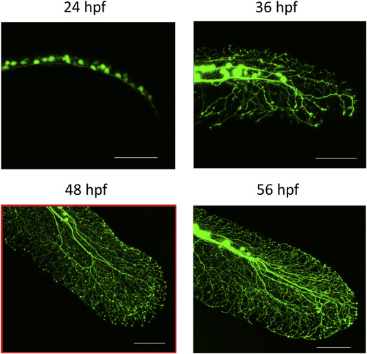
Fig. 1 Sensory neurite development in the caudal fin of sensory:GFP embryos at different timepoints. Sensory neurite development in the caudal fin of sensory:GFP embryos was followed at several stages of development (24, 36, 48, and 56 hpf). Indicated by the red frame is the developmental stage (48 hpf) that was used to study the effect of the pathogenic p.(I228M) and p.(G856D) mutations. Images represent maximal projections of confocal recordings at 20× magnification. Scale bar indicates 100 μm.
Figure Data
Acknowledgments
This image is the copyrighted work of the attributed author or publisher, and
ZFIN has permission only to display this image to its users.
Additional permissions should be obtained from the applicable author or publisher of the image.
Full text @ Exp. Neurol.