
Fig. 3
- ID
- ZDB-IMAGE-190201-29
- Genes
- Antibodies
- Publication
- Sokol et al., 2018 - Loss of the Mia40a oxidoreductase leads to hepato-pancreatic insufficiency in zebrafish
- All Figures
- Figures for Sokol et al., 2018
|
Fig. 3
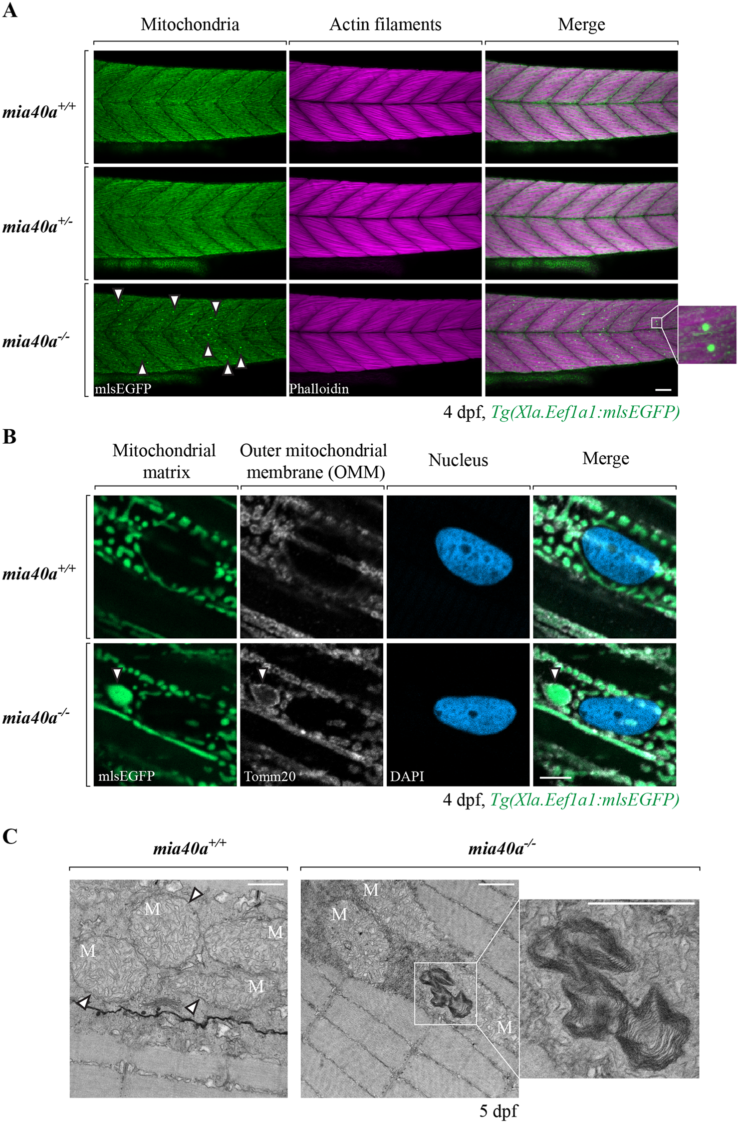
Abnormal mitochondrial structures are found in the skeletal muscles of mia40a mutants.
(A) Embryos obtained from an in-cross of heterozygous mia40a+/- siblings in the Tg(Xla.Eef1a1:mlsEGFP) background were stained with phalloidin to visualize actin filaments of the skeletal muscle at 4 dpf. Abnormal, enlarged mitochondrial structures are found in the skeletal muscles of the mia40a mutants (arrowheads). To better visualize the GFP-positive inclusions, a magnified image is shown on the side. Images are maximum projections. Scale bar, 50 μm. All images are lateral views, anterior to the left. (B) wild-type and mia40a-/- larvae in the Tg(Xla.Eef1a1:mlsEGFP) background were stained with an anti-Tomm20 antibody to mark the outer mitochondrial membrane (OMM). DNA was counterstained with DAPI. Single plane sections are shown. The arrowhead points to a GFP-positive inclusion. Scale bar, 5 μm. All images are lateral views, anterior to the left. (C) wild-type or mia40a-/- siblings in AB background were subjected to transmission electron microscopy (TEM) analysis at 5 dpf. Arrowheads point to well defined mitochondrial membranes in wild-type larvae. Abnormal membranous structures found in the mia40a mutants are magnified (inset). Scale bar, 500 nm. M: Mitochondria.