
Image
|
Figure Caption
Fig. 3
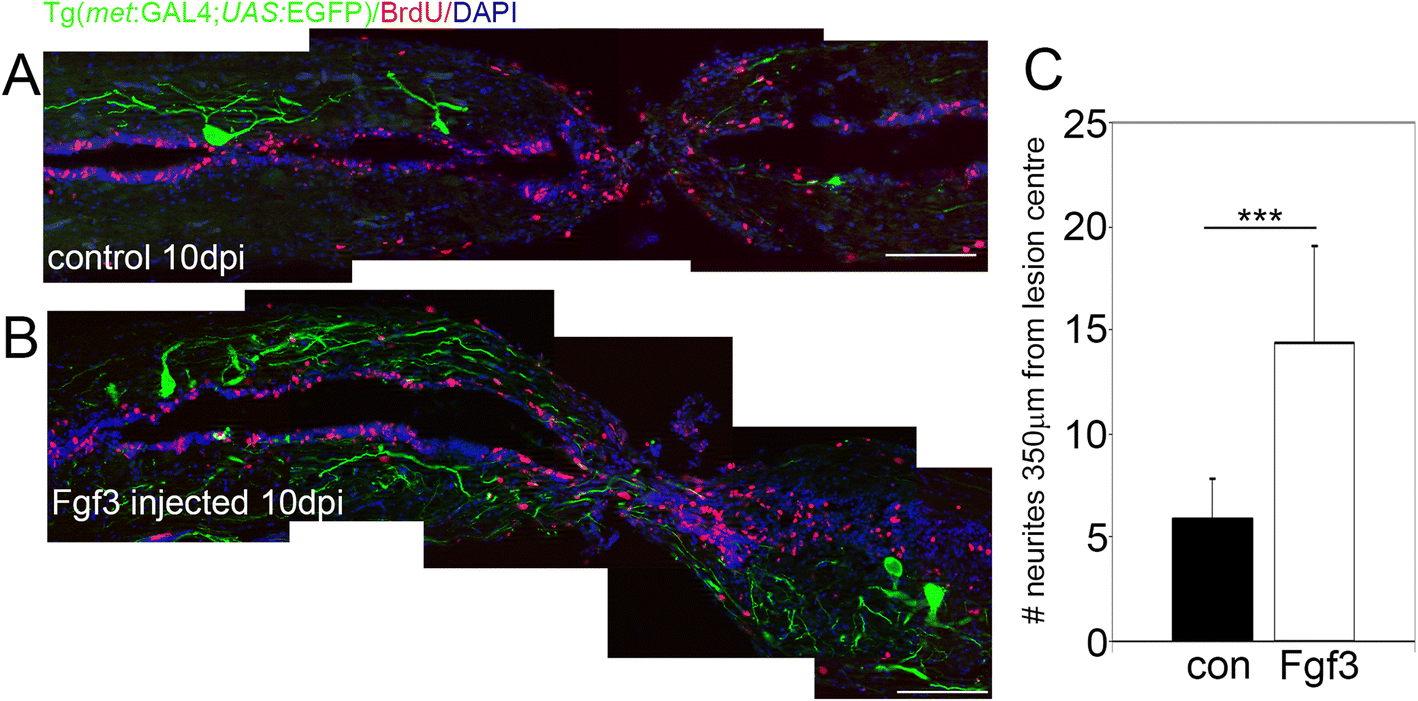
Fgf3 facilitates neurite sprouting of C-Met motor neurons after spinal cord injury. a, b Longitudinal sections through the spinal cord lesion site reveal that ten days post injury (dpi) Fgf3 treatment resulted in significantly more neurites at the lesion. Scale bars in A and B are 200 μm. c Quantitation of neurites up to 350 μm from lesion centre from both sides. Results are presented in C as mean ± SEM, (n = 7 fish /group) *** p < 0.001
Acknowledgments
This image is the copyrighted work of the attributed author or publisher, and
ZFIN has permission only to display this image to its users.
Additional permissions should be obtained from the applicable author or publisher of the image.
Full text @ Neural Dev.